
Method for substantially non-delaminable smooth ventricular assist device conduit and product from same

a technology of ventricular muscle and assist device, which is applied in the direction of catheters, blood vessels, therapy, etc., can solve the problems of ventricular muscle over-stretches, high cost associated with the condition, and shortness of breath, so as to achieve non-thrombogenic porous, not safe for the patient, and high cost. the effect of the condition

Active Publication Date: 2010-01-05
EDWARDS LIFESCIENCES CORP
View PDF13 Cites 0 Cited by
  • Summary
  • Abstract
  • Description
  • Claims
  • Application Information

AI Technical Summary

Benefits of technology

[0028]Accordingly, it is an object of the present invention to provide a method of substantially non-delaminably bonding a substantially non-porous thermoplastic elastomer to a substrate. A further object of the present invention is to provide a substantially non-delaminable conduit for LVAD's that has a highly non-thrombogenic porous inner surface and is not susceptible to air leakage under negative pressure. Another object of the present invention to provide a substantially non-delaminable conduit for LVAD's that has a highly non-thrombogenic porous inner surface, is not susceptible to air leakage under negative pressure, and is relatively safe for the patient. Still another object of the present invention is to provide a substantially non-delaminable conduit for LVAD's, wherein said conduit has a highly non-thrombogenic porous inner surface, is not susceptible to air leakage under negative pressure, and is relatively easy to use. An additional object of the present invention is to provide a substantially non-delaminable conduit for LVAD's that is comparatively cost-effective to manufacture. Even still a further object of the present invention is to provide a substantially non-delaminable conduit for LVAD's that has a highly non-thrombogenic porous inner surface, is not susceptible to air leakage under negative pressure, and is supported by a substantially non-delaminable support structure. Yet still a further object of this invention is to provide methods and apparatus that are suitable for use with the variety of polymeric materials that are used in LVAD devices. Even still another object of this invention is to provide a LVAD apparatus that allows the surgical practitioner to operate the apparatus in the open chest of the patient during the implantation procedure. Even yet still another object of this invention is to provide a method for the treatment of congestive heart failure using a LVAD apparatus that allows the surgical practitioner to operate the apparatus in the open chest of the patient during the implantation procedure. Even an additional object of this invention is to provide an article of manufacture for packaging the apparatus of the invention.

Problems solved by technology

The high costs associated with the condition will place an added burden on public health resources as the incidence of CHF continues to rise.
This can result in a shortness of breath, fatigue and fluid retention (edema) and if left unchecked can lead to death within a few years.
As the condition worsens, the ventricular muscle over-stretches and the muscle fails to work to its full efficiency.
This leads to a further reduction in the cardiac output and exacerbates symptoms of heart failure.
The combined effect of these responses can lead to the formation of edema, especially in the legs and ankles.
If heart failure occurs in the left side of the heart, pulmonary edema can result which manifests as breathlessness.
In advanced CHF, the severity of the symptoms can be disabling and often leads to hospitalization.
The failing heart is a result of a number of factors combining to reduce the efficiency of the heart as a pump.
As the blood flow from the heart slows, the blood returning to the heart through the veins can back-up, resulting in congestion in the tissues.
This can lead to swelling in the legs and ankle and fluid retention in the lungs, which interferes with breathing and contributes to the characteristic shortness of breath seen in people with CHF.
The demands on artificial blood conduits in ventricular assist devices are great.
Moreover, there are differences in flow and pressure between the inflow and outflow conduits connected to the pumping device.
However, such conduits alternately collapsed and regained their tubular shape with each stroke of the pump.
Prior art improvements comprised creating a scaffolding which prevented the collapse of the conduit with each pump stroke. however, since the tube no longer collapsed, the lumen of the tube experienced negative pressure and atmospheric air consequently infiltrated through the walls into the lumen.
Porosity also renders an inflow conduit vulnerable to the entrance of air during the intervals of negative pressure transients that can occur in the inflow conduit as noted above.
This raises a serious problem whenever the inflow conduit is in contact with the atmosphere.
Air can be sucked into the inflow conduit during intervals of negative pressure transients as outlined above, leading to air embolisms in the cardiovascular system of the patient undergoing the implantation procedure.
Such air embolisms can lead to injury to the patient or even to the death of the patient.
It is obvious, therefore, that this risk of air embolisms resulting from the vulnerability of the inflow conduit to the entrance of air during intervals of negative pressure, whenever the inflow conduit is in contact with the air, is an extremely serious problem.
Even if the conduits are sealed with a thin bio-compatible collagen coating on the inner lumen wall to render the fabric more leak resistant at the time of implantation this procedure only addresses the problem of leakage of the lumen contents outward, and does not at all address the problem of the entrance of air during the intervals of negative pressure transients that can occur in the inflow conduit as noted above.
Such conduits might solve the problem of leakage into the conduit lumen when it is under negative pressure, but they would not be suitable for use in implantable devices, as they will not easily accommodate varying anatomical placements, and tend to kink if bent.
Thus, even this device does not solve the twin requirements of a conduit having a highly non-thrombogenic porous inner surface that is not susceptible to leakage under negative pressure.
Even though conduits for LVAD's are used extensively in medical practice, prior devices, products, or methods available to medical practitioners have not adequately addressed the need for non-delaminable apparatus for conduits for LVAD's to meet the need for methods and apparatus for conduits for such pumps as outlined above.

Method used


Image

  • Method for substantially non-delaminable smooth ventricular assist device conduit and product from same
  • Method for substantially non-delaminable smooth ventricular assist device conduit and product from same
  • Method for substantially non-delaminable smooth ventricular assist device conduit and product from same

Examples


Embodiment Construction

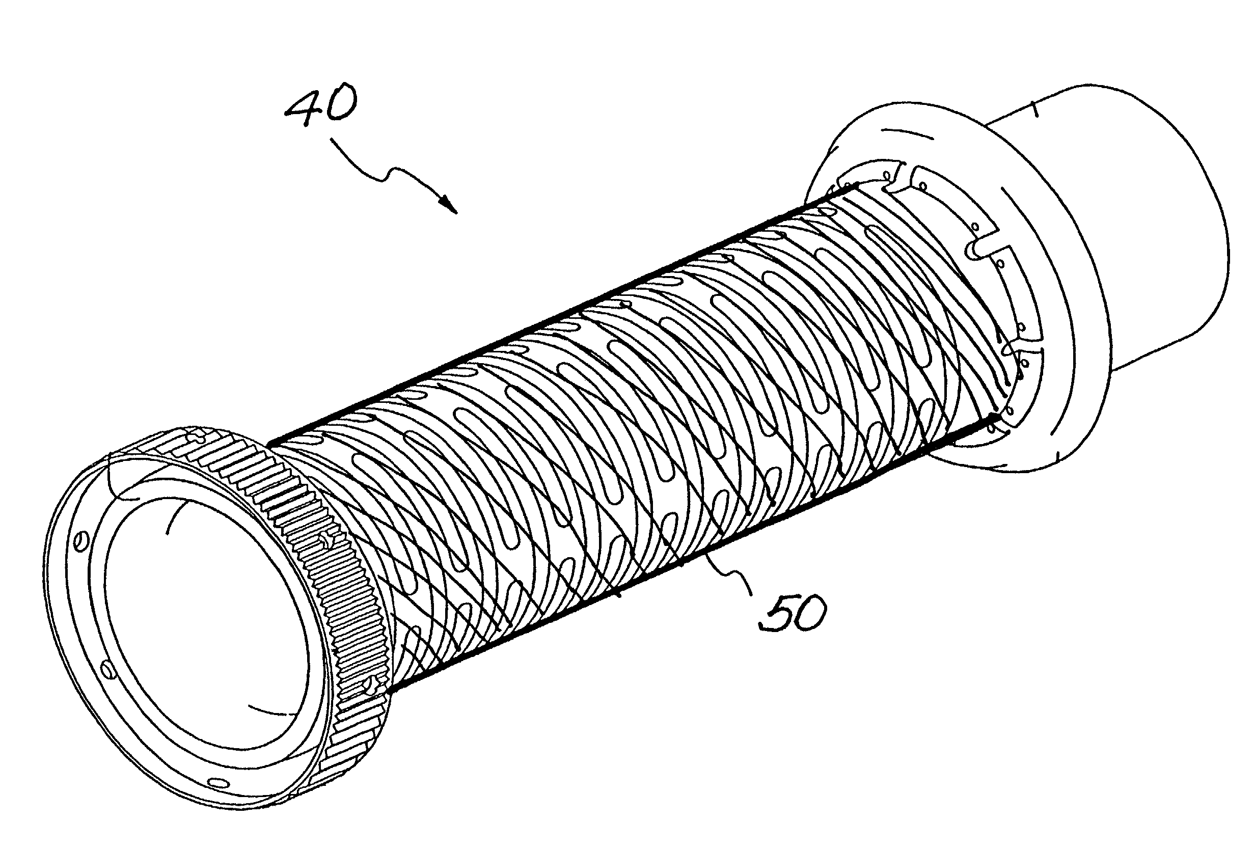
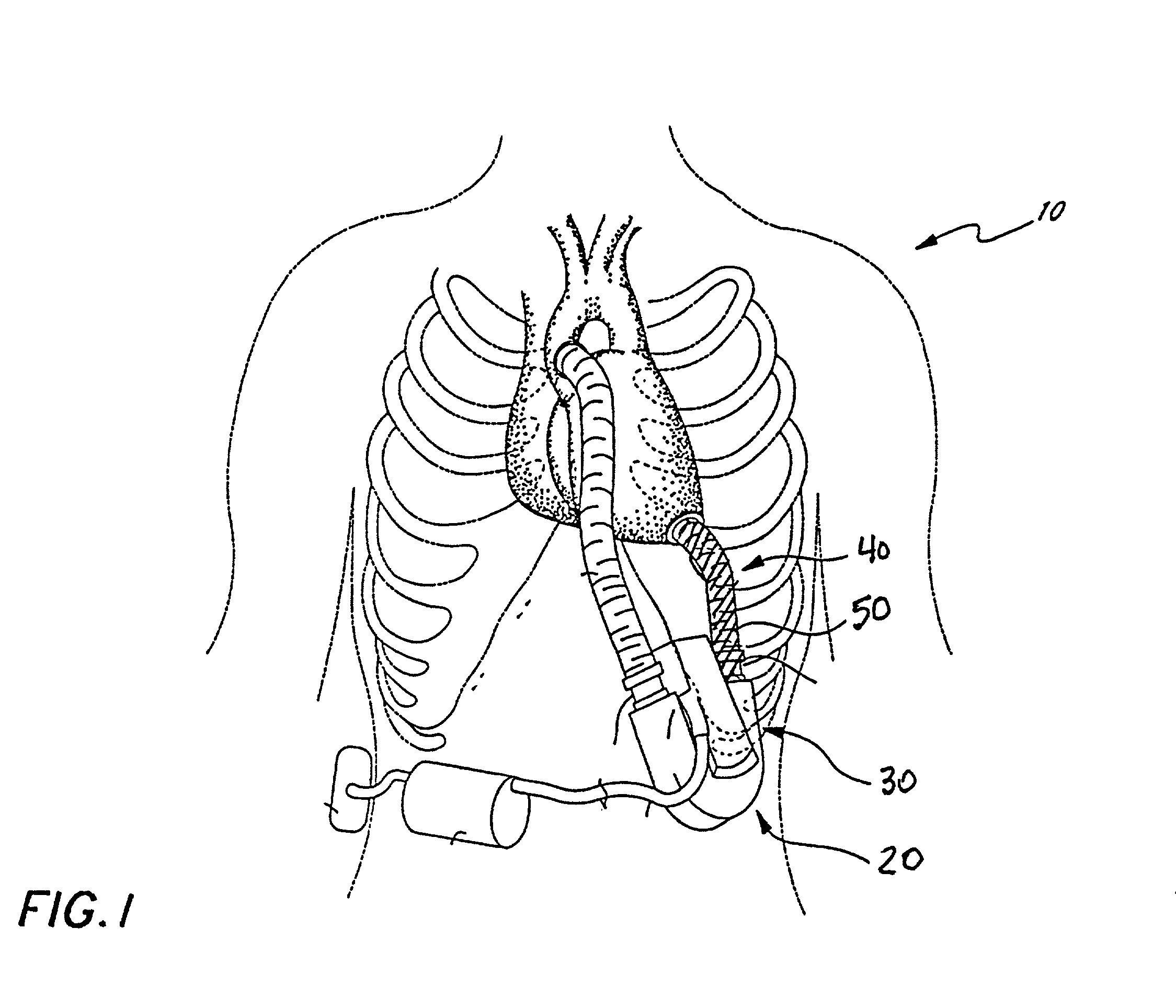
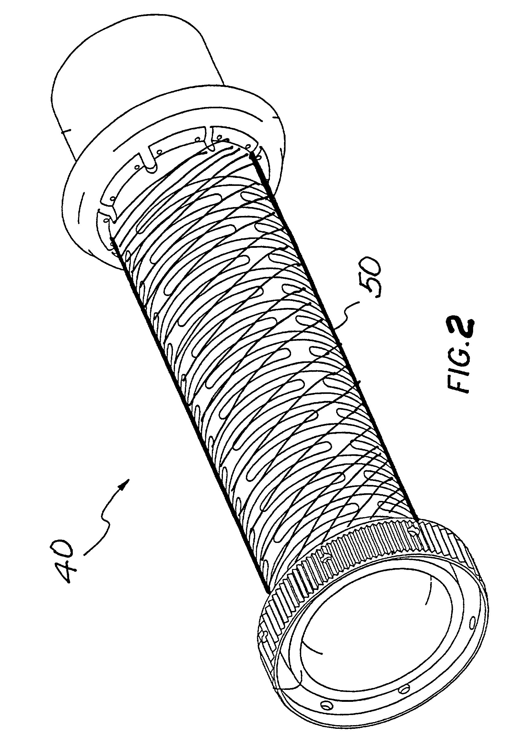
[0034]With reference to FIGS. 1 and 2, a living human host patient 10 with an open chest is shown in fragmentary front elevational view, and with parts of the patient's anatomy shown in phantom or removed solely for better illustration of the salient features of the present invention. Surgically implanted into the patient is the pumping portion 20 of a ventricular assist device, generally referenced with the numeral 30. Ventricular assist device 30 includes an inflow conduit 40 which further includes a substantially non-porous conduit 50 for communicating blood from the patient's left ventricle into the pumping portion 20. As shown in FIG. 2, conduit 50 comprises a flexible, porous tubular substrate to which a substantially non-porous thermoplastic elastomer is substantially non-delaminably bonded in accordance with a method of the invention. An end of inflow conduit 40 is connected to the patient's heart by sutures so that blood flow communication is established and maintained.

[003...


PUM

PropertyMeasurementUnit
water entry pressureaaaaaaaaaa
pressureaaaaaaaaaa
thicknessaaaaaaaaaa

Abstract

A method of non-delaminably bonding a non-porous thermoplastic elastomer to a substrate by heating a bilayer of the elastomer and the substrate for a predetermined time and a product obtained by the method. Methods for producing non-delaminable conduits and non-delaminable kink-resistant conduits constructed of biocompatible elastomers and substrates, and products produced by these methods are taught. Methods, products, and articles of manufacture relating to non-delaminable monofilament supported kink-resistive conduits that may be used as inflow conduits in left ventricular assist devices for treatment of heart failure are also provided.

Description

BACKGROUND ART[0001]The present invention is related to methods and apparatus for surgically implantable pumps that provide a mechanical device for augmenting or replacing the blood pumping action of damaged or diseased hearts. More specifically, this invention is related to methods and apparatus for conduits for such pumps to meet the three-fold requirements of a substantially non-delaminable conduit having a highly non-thrombogenic porous inner surface that is not susceptible to air leakage under negative pressure, whereby the use of such implantable pump procedures is broadly enabled, and wherein the functional utility, ease of use, and wide applicability of the device in medical practice constitutes progress in science and the useful arts. Furthermore, the present invention teaches processes to make and use the device in medical practice.[0002]Of all the cardiovascular disorders, congestive heart failure (CHF) is the only one to show a sharp increase in prevalence since the 1960...

Claims


Application Information

Patent Timeline
Patent Type & Authority Patents(United States)
IPC IPC(8): A61M25/00A61L29/04A61L29/08A61L31/04A61L31/10A61M60/178A61M60/205A61M60/857B32B1/08
CPCA61L29/041A61L29/085A61L31/048A61L31/10A61M1/10B32B1/08B32B37/04C08L27/18A61M1/122B32B2327/18A61M60/148A61M60/178A61M60/205
Inventor SHANNON, DONALD T.KUO, CHRISTU, BENITARANDALL, MIKE
Owner EDWARDS LIFESCIENCES CORP
Learn More