
Cultured cartilage/bone cells/tissue, method of generating same and uses thereof

a technology of cartilage and bone cells, which is applied in the field of cultured cartilage/bone cells/tissue, method of generating same and uses thereof, can solve the problems of not being aware of their existence nor function, damaged cartilage tissue does not receive sufficient or proper stimuli to elicit a repair response, and arthritis alone is an enormous medical and economic problem

Inactive Publication Date: 2010-01-05
TECHNION RES & DEV FOUND LTD
View PDF6 Cites 20 Cited by
  • Summary
  • Abstract
  • Description
  • Claims
  • Application Information

AI Technical Summary

Benefits of technology

The present invention provides an isolated mandibular condyle tissue with a reduced amount of fibroblast-like cells and myocytes, and a method of isolating and culturing chondrocytes from this tissue. The isolated chondrocytes can be used for treating cartilage and bone diseases in a subject by administering a therapeutically effective dose of the cultured chondrocytes. The culturing conditions used for the isolated chondrocytes include devoid of a three-dimensional support and / or a biomolecule-coated support. The invention also provides a cell culture containing the isolated chondrocytes and a method of redifferentiating dedifferentiated chondrocytes.

Problems solved by technology

Failure of diarthrodial joints leads to arthritis, the most common form being osteoarthritis.
Arthritis alone is an enormous medical and economic problem, with more than thirty million Americans suffering from this highly debilitating disease.
They perform their function so well that we are often not even aware of their existence nor the function they provide until injury strikes or arthritis develops.
It is generally believed that because articular cartilage lacks a vasculature, damaged cartilage tissue does not receive sufficient or proper stimuli to elicit a repair response.
Damage to these structures can lead to a reduction in joint function and degeneration of the articular cartilage, and surgical removal of such damaged structures, usually the main treatment, can result in early onset of osteoarthritis.
The structure of proteoglycan in cartilage is not uniform.
Second, evidence of strong frictional interaction between the proteoglycans and the fine collagen network also exists.
Degenerative loss of articular cartilage, for example, at the acetabular / femoral head articulation, results in heavy loading of the soft tissue, and can require radical surgery.
There are significant limitations with all present approaches.
Artificial prostheses have a limited lifetime, and can fail prematurely.
Recurrent replacements of prostheses is not an advisable treatment, and, therefore, the relatively young and active patient is often consigned to slow joint degeneration until the use of prosthetic implants becomes a reasonable clinical option.
Prostheses rarely replicate the performance of the original tissue.
A prosthesis cannot adapt in response to environmental stress as does a biological tissue, nor can it repair itself.
Biological allograft material is in limited supply, appropriate size shape and tissue type are difficult to obtain, and has the risk of carrying infectious diseases.
Use of autograft material compromises the site used for the source tissue (e.g., using patella tendon to replace anterior cruciate ligament), and can only offer this tissue once.
However, culturing cartilage / bone from primary chondrocytes has been found to be highly problematic due to the fact that primary cartilage-derived cell cultures undergo dedifferentiation, acquire fibroblastic features, and lose most of the characteristics of mature chondrocytes.
This dedifferentiation phenomenon is furthermore a serious obstacle for ex-vivo studies of the endochondral ossification process and its extra- and intracellular regulation, and for in-vitro studies of various articular pathologies such as rheumatoid arthritis or osteoarthritis.
All of the aforementioned approaches, however, suffer from significant disadvantages including: incapacity to generate cultured cartilage / bone displaying optimal cartilage / bone specific differentiation, and / or displaying such differentiation for an optimally long duration in-vitro; and / or their being excessively cumbersome / complex and / or expensive to practice, such as in the case of approaches involving the use of three dimensional supports or biomolecule-coated substrates.
Thus, all prior art approaches have failed to provide an adequate solution for generating cultured cartilage / bone.

Method used


Image

  • Cultured cartilage/bone cells/tissue, method of generating same and uses thereof
  • Cultured cartilage/bone cells/tissue, method of generating same and uses thereof
  • Cultured cartilage/bone cells/tissue, method of generating same and uses thereof

Examples


example 1

Method of Optimally Isolating Mandibular Condyle Chondrocytes, and of Culturing Primary Chondrocytes to Optimally Generate Optimally Differentiated Cultured Chondrocytes / Endochondral Bone Cells

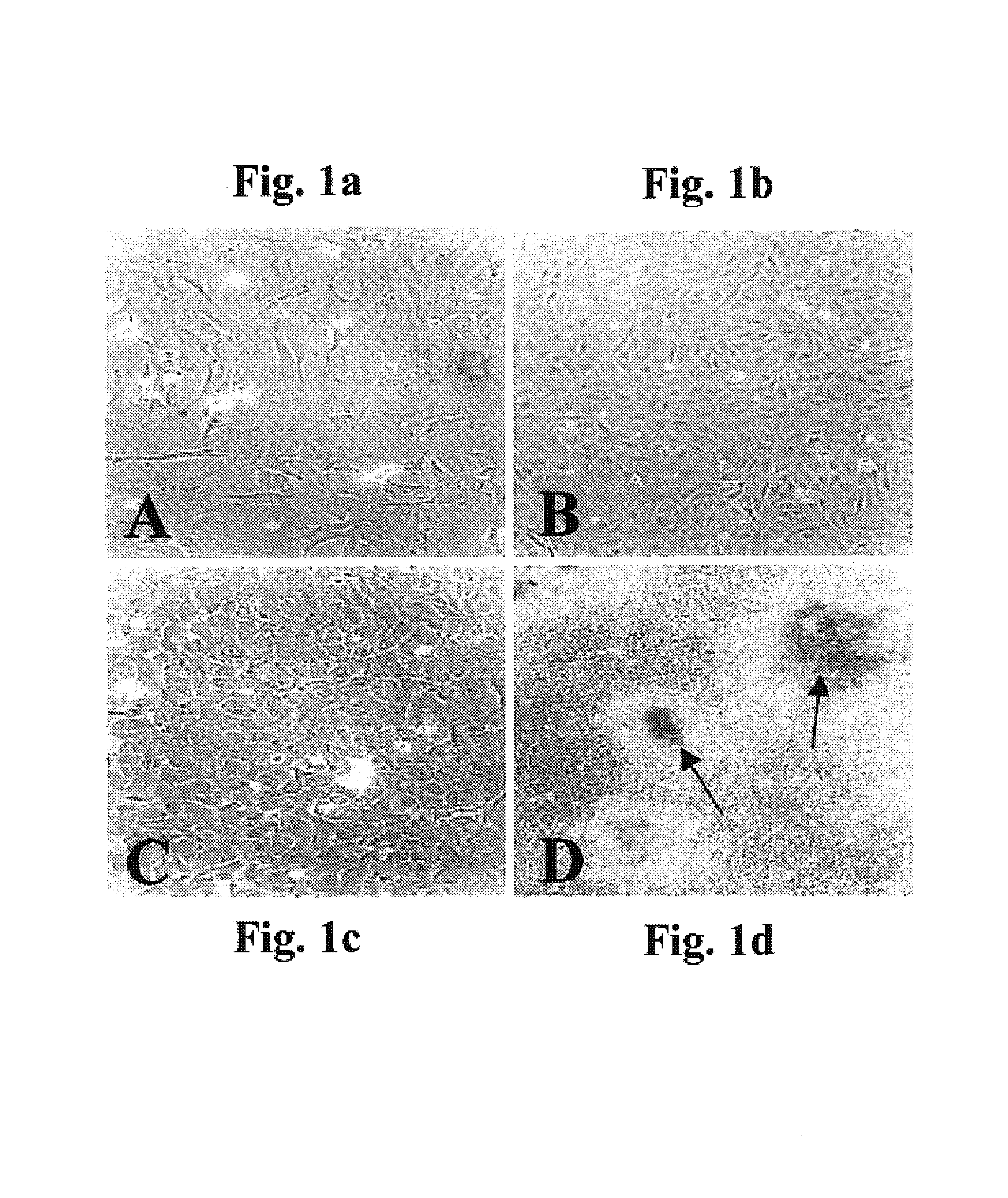
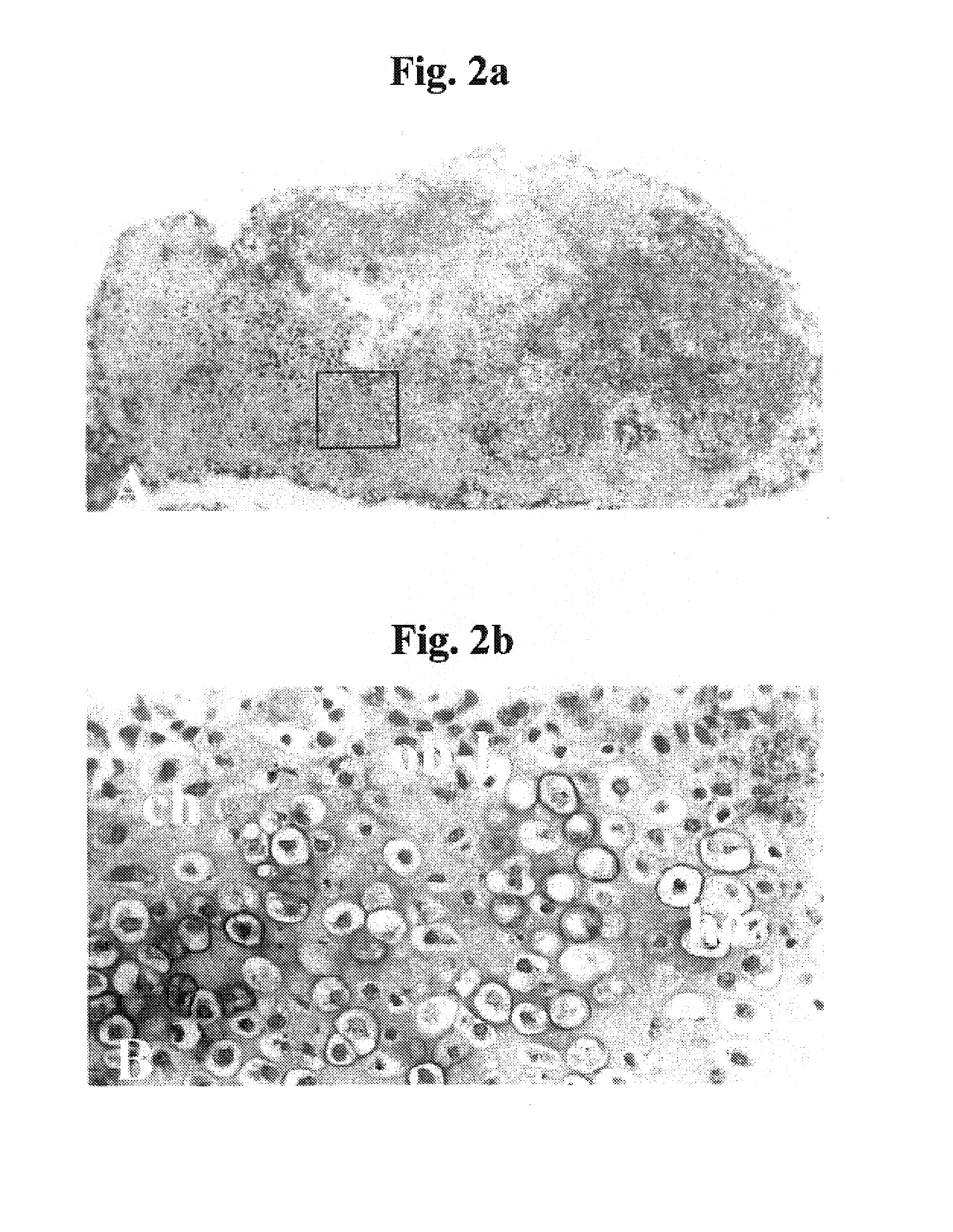
[0238]Background: Cartilage / bone diseases include numerous highly debilitating diseases, such as arthritis, which are of tremendous medical and economic impact and for which no optimal treatment methods exist. An optimal strategy for treating such diseases would be to generate and administer cultured chondrocytes / endochondral bone cells so as to replace / repair lost / damaged cartilage / bone. While various methods of generating cultured chondrocytes / bone have been proposed in the prior art, these have been highly suboptimal for various reasons, as described in the Field and Background of the Invention section above. Notably, primary chondrocyte-derived cell cultures tend to undergo dedifferentiation, acquire fibroblastic features, and lose most of the characteristics of mature chondrocytes, and th...

example 2

Genetic Transformation of Isolated Mandibular Condyle Derived Chondrocytes

[0266]Materials and Methods:

[0267]The capacity to genetically transform cultured mandibular condyle derived chondrocytes (MCDCs) isolated as described in Example 1, above, would be potentially highly useful in therapeutic applications involving cell therapy with such cells and for investigating chondrocyte biology in-vitro.

[0268]Constructs: Sequences encoding osteoprotegerin (OPG) were cloned into expression vector pIRES2-EGFP (Clontech) yielding the bicistronic expression vector OPG-IRES-GFP for co-expression of osteoprotegerin and green fluorescent protein (GFP) to thereby generate expression construct pcDNAI-OPG-IRES-GFP. Sequences encoding a fusion protein composed of GFP fused to glucose transporter GLUT4 were cloned into expression vector pcDNAneoI to thereby generate expression vector pcDNAI-GLUT4-GFP.

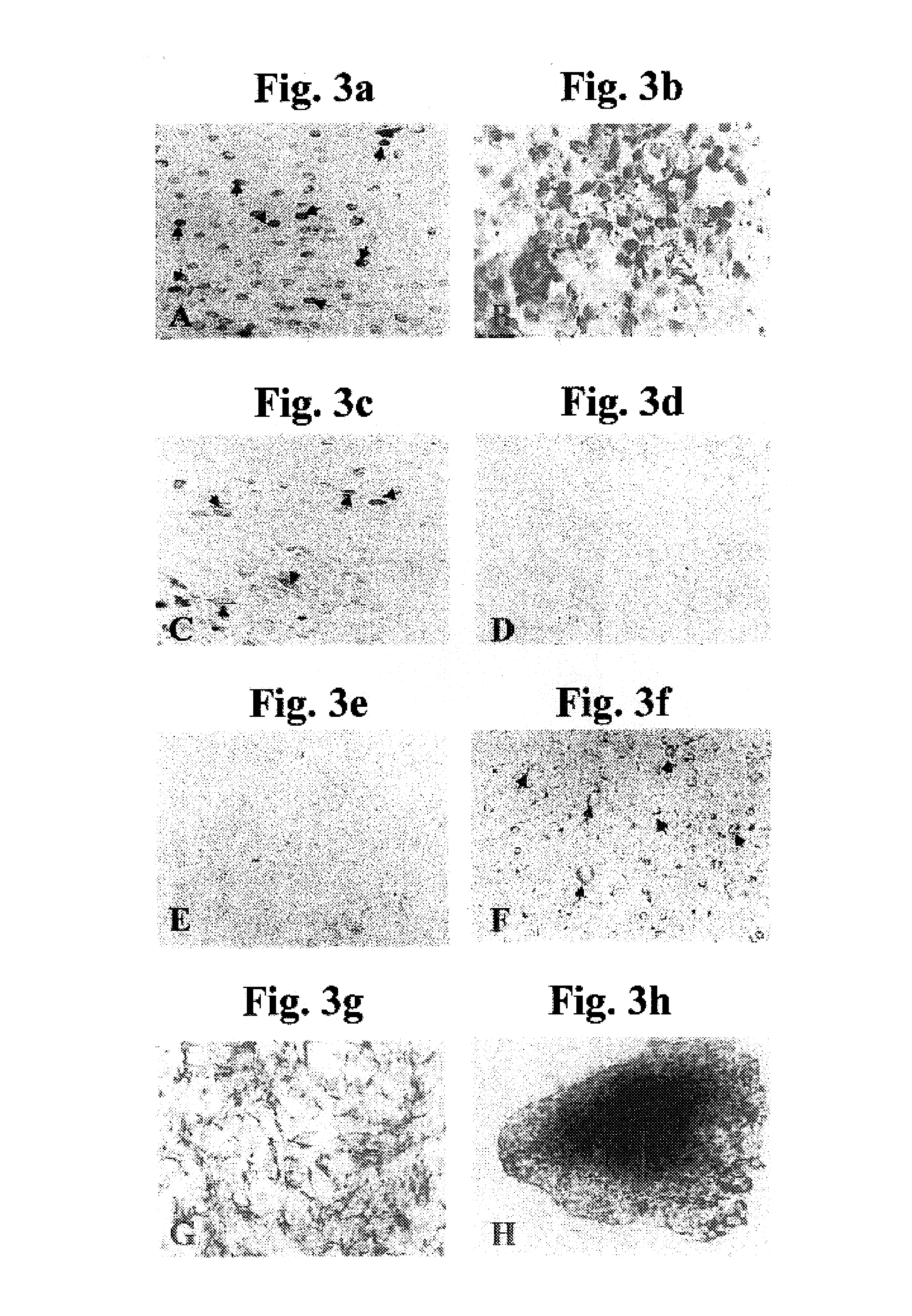
[0269]Transfection of cultured mandibular condyle derived chondrocytes: MCDCs were cultured in 24-well ...


PUM

PropertyMeasurementUnit
molecular weightsaaaaaaaaaa
lengthaaaaaaaaaa
molecular weightaaaaaaaaaa

Abstract

A method of generating cultured chondrocytes / endochondral bone cells is provided. The method comprising isolating chondrocytes from mandibular condyle tissue, and culturing the isolated chondrocytes. A method of isolating chondrocytes from mandibular condyle tissue is further provided. The method comprises isolating mandibular condyle tissue from a mammal and treating the mandibular condyle tissue so as to selectively remove fibroblast-like cells and / or myocytes therefrom, the modified mandibular condyle tissue including chondrocytes, and selectively collecting the chondrocytes from the modified mandibular condyle tissue.

Description

[0001]This application claims the benefit of priority from U.S. Provisional Patent Application No. 60 / 450,688, filed Mar. 3, 2003, the contents of which are hereby incorporated by reference.FIELD AND BACKGROUND OF THE INVENTION[0002]The present invention relates to methods of isolating chondrocytes, methods of culturing isolated chondrocytes to generate cultured chondrocytes / bone cells, and to uses of such cultured chondrocytes / bone cells. More particularly, the present invention relates to methods of isolating mandibular condyle chondrocytes, methods of culturing same to generate highly differentiated cultured chondrocytes / endochondral bone cells, and to uses of such cultured chondrocytes / endochondral bone cells for treating cartilage / bone diseases.[0003]Cartilage / bone diseases include highly debilitating and / or lethal diseases such as arthritis, articular cartilage injury, meniscal disorders, joint infections, chondrogenesis disorders and cosmetic disorders of cartilaginous struct...

Claims


Application Information

Patent Timeline
Patent Type & Authority Patents(United States)
IPC IPC(8): C12N5/00A61K35/12C12N5/077
CPCC12N5/0655C12N2501/105A61K35/12C12N2500/38A61P19/00
Inventor MAOR, GILA
Owner TECHNION RES & DEV FOUND LTD
Learn More