Method of bioimage data processing for revealing more meaningful anatomic features of diseased tissues

a bioimage and data processing technology, applied in image enhancement, instruments, applications, etc., can solve the problems of abnormal thickness of retina, abnormal overall contour of retina, and thin retina

Active Publication Date: 2010-02-23
CARL ZEISS MEDITEC INC
View PDF10 Cites 37 Cited by
  • Summary
  • Abstract
  • Description
  • Claims
  • Application Information

AI Technical Summary

Problems solved by technology

In ophthalmology, retinal thickness may be abnormally large in cases of retinal edema or traction by membranes in the vitreous humor.
On the other hand, the retina may appear thin in cases of atrophic degeneration, chorioretinitis, or trauma to the retina.
In certain cases, the overall contour of the retina may become abnormal.
However, a major issue associated with a tissue layer thickness map is that it sometimes cannot reveal the diagnostically more meaningful features of a diseased part of the tissue.
As a result, a retina thickness map such as the color coded one shown in FIG. 2, which corresponds to FIG. 1, cannot reveal the substantially raised bump.
On the other hand, although a topographic map or contour of the RPE or ILM may reveal the sharp bump better for this illustrated case than the retina thickness map, it would include both the sharp bump and the broader warping of the RPE boundary, making it difficult to separate the effect of the disease from the overall shape of the RPE boundary

Method used


Image

  • Method of bioimage data processing for revealing more meaningful anatomic features of diseased tissues
  • Method of bioimage data processing for revealing more meaningful anatomic features of diseased tissues
  • Method of bioimage data processing for revealing more meaningful anatomic features of diseased tissues

Examples


Embodiment Construction

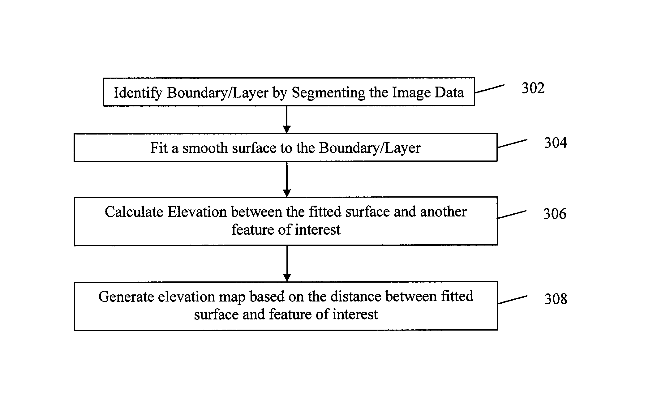
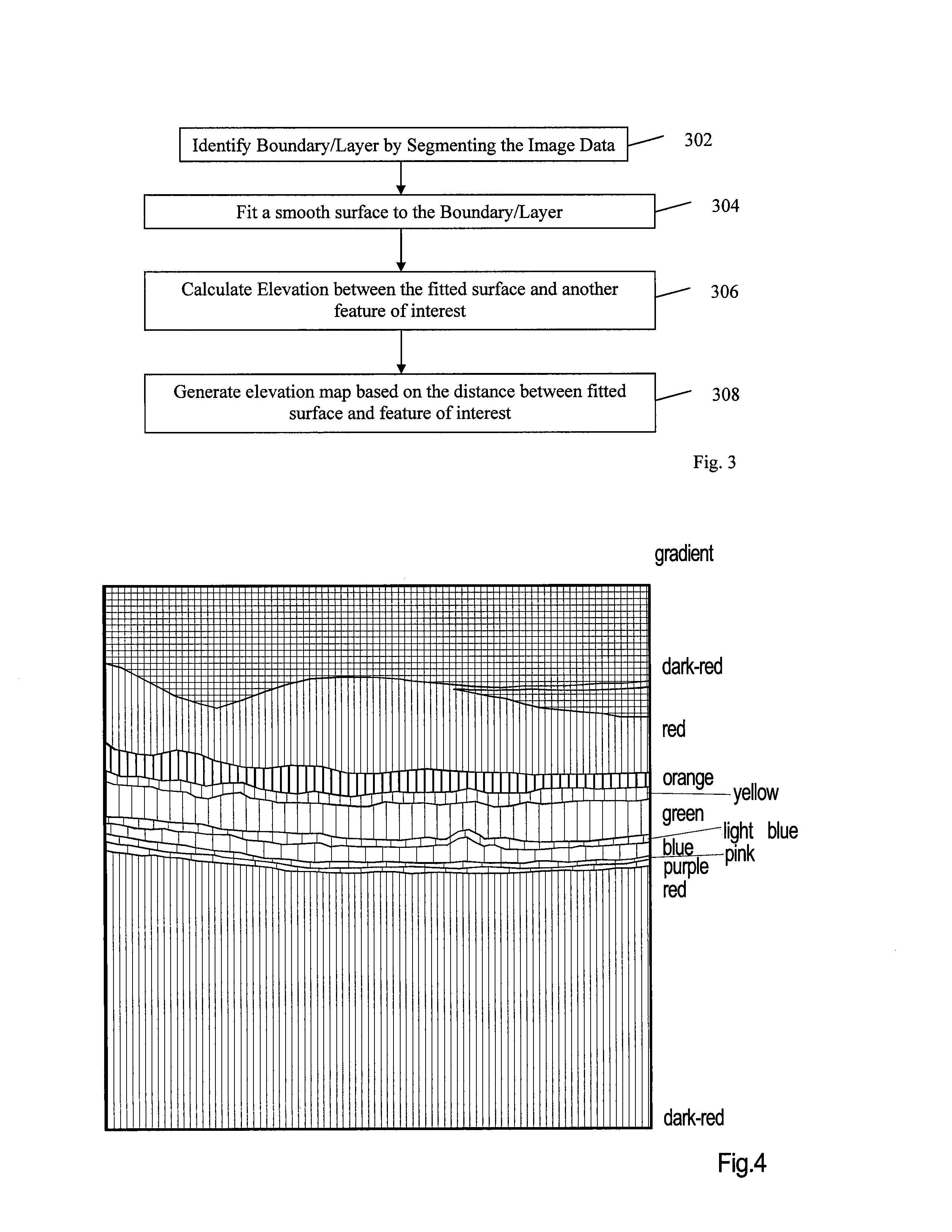
[0026]FIG. 3 shows one preferred embodiment the presently invented method. This method is intended to be used on image data obtained from a sample. The illustrations in this application are based on image data derived from an optical coherence tomography system (OCT) which includes both time domain and spectral domain OCT systems. Such an instrument generates 3D intensity data corresponding to an axial reflection distribution arising from reflecting features in the eye. As noted above, this information is currently used by doctors to view and diagnosis various pathologies in the eye. A basic OCT system will be discussed below.

[0027]Although the illustrated embodiments are limited to OCT data, the image processing concepts described herein may be used with 3D image data derived from other modalities. For example, image data may be created using various forms of confocal microscopy and even ultrasound imaging modalities.

[0028]The first step in the subject method requires identificatio...


PUM


Abstract

The present invention discloses a method for generating elevation maps or images of a tissue layer / boundary with respect to a fitted reference surface, comprising the steps of finding and segmenting a desired tissue layer / boundary; fitting a smooth reference surface to the segmented tissue layer / boundary; calculating elevations of the same or other tissue layer / boundary relative to the fitted reference surface; and generating maps of elevation relative to the fitted surface. The elevation can be displayed in various ways including three-dimensional surface renderings, topographical contour maps, contour maps, en-face color maps, and en-face grayscale maps. The elevation can also be combined and simultaneously displayed with another tissue layer / boundary dependent set of image data to provide additional information for diagnostics.

Description

TECHNICAL FIELD OF THE INVENTION[0001]One or more embodiments of the present invention relate generally to methods for optical imaging of biological samples and for processing such images. In particular, the invention is a method for processing a three-dimensional image data set to generate elevation maps of tissue layers relative to a fitted smooth surface, which can provide more diagnostic information than a pure tissue layer thickness map. Maps of elevation may be embodied as three-dimensional surface renderings of elevation, topographical maps, or as color or grayscale maps.BACKGROUND OF THE INVENTION[0002]Measurement of biological tissue surface contour or layer thickness can provide useful diagnostic information in various applications. For example, arterial plaque thickness is related to the progress of atherosclerosis, carotid vessel wall thickness is also an indicator of cardiovascular disease risk; epidermal layer thickness is an indicator of burn severity.[0003]In ophthal...

Claims


Application Information

Patent Timeline
Patent Type & Authority Patents(United States)
IPC IPC(8): G06K9/00A61B5/103G01B9/02
CPCG06T19/00A61B5/0066G06T7/0014G06T2210/41G06T2207/30041G06T2200/24G06T2207/10101
Inventor EVERETT, MATTHEW J.MEYER, SCOTT A.STETSON, PAUL F.ZHOU, YAN
Owner CARL ZEISS MEDITEC INC
Why Eureka
Try Eureka