Breakaway interfacing of radiological images with work orders

a work order and radiological image technology, applied in the field of computer-aided imaging systems and computer-aided radiological information systems, can solve the problems of inability to accurately assess the accuracy of radiological images, the slice may not be evenly distributed across the patient's body, and the machine is a very expensive asset, so as to reduce the need for human intervention, improve the effect of highly automated communication, and reduce the likelihood of errors

Inactive Publication Date: 2010-07-13
DEJARNETTE RES SYST
View PDF5 Cites 6 Cited by
  • Summary
  • Abstract
  • Description
  • Claims
  • Application Information

AI Technical Summary

Benefits of technology

[0015]The present invention provides, for use in combination with medical imaging equipment normally operating independently of a stand-alone radiological information system, a breakaway interface disposed between the radiological information system and PACS and the medical imaging equipment. This breakaway interface facilitates conventional use of the medical imaging equipment for multi-anatomical or multi-procedural studies for generating a series of anatomical images under a single work order and for simultaneously producing respective individual work orders which are matched to corresponding anatomical images, and which are inputted into the radiological information system for management control, tracking, accounting and / or billing purposes.
[0020]A first object of the invention is to provide improved and more highly automated communication between a radiological information system and a radiological imaging machine. A second object of the invention is to reduce the need for human intervention and interpretation, and consequently reduce the likelihood for errors. A third object of the invention is to ensure that appropriate information is stored with each appropriate work order, whereby at a later date the imaging information may be readily obtained for review and comparison. A fourth object of the invention is to permit both a radiological information system and a radiological imaging machine to be operated using native logic and associated work orders, whereby the benefits of each system are preserved and allowed to remain optimal for their intended purposes. Another object of the invention is to provide filtered conversions both from a radiological information system to a radiological imaging machine and in an opposite direction from said radiological imaging machine to said radiological information system. A further object of the invention is to maintain compliance with existing standards, such as the DICOM standards. These and other objects are achieved in the present invention, which may be best understood by the following detailed description and drawing of the preferred embodiment.

Problems solved by technology

These machines are very expensive assets that will most desirably be operated in an efficient manner to minimize idle time and, simultaneously, to minimize the amount of time required to produce a desired set of images for a given patient.
These slices may not be evenly distributed across a patient's body, but in accord with a physicians' needs may be unevenly concentrated in a particular body area or region.
Unfortunately, the objectives of the various systems are somewhat different from each other, and, as a result, certain technical challenges are present in the integration of the various components that comprise a complete radiological program.
Unfortunately, when there is a multi-anatomical study or multi-procedure study work order placed, which is very well handled by many axial medical imaging scanners, neither the PACS system nor the radiological information system are well suited.
Consequently, for a multi-anatomical study, the remaining image studies could not be assigned by the reading radiologist, and these studies remained unassigned and not associated with any particular work order.
As a result, the facility was unable to properly account, track and bill for the remaining image studies.
The work orders, however, indicate three separate procedures, a discrepancy that may, particularly in the case of a new or less-experienced technician, lead to error, confusion or delay, any of which is associated with a decreased level of service and the potential need to expose the patient multiple times to the imaging radiant energy.
When the CT technician properly completes the single comprehensive procedure, yet another problem exists.
In either case, additional work not associated directly with patient care is undesirably required.
In the event the multi-study imaging output is not divided (which is tedious work not directly affiliated with patient care) issues arise regarding the accounting between the radiology report and the radiological information system.
When work orders are properly divided, but the images are not, the remaining work orders are all too frequently orphaned, unmatched to any image set in spite of the fact that the imaging study has actually been completed.
In either case, when work orders are not properly divided or images are not properly divided, another very significant benefit of the PACS is lost.
Unfortunately, when either the work order or the imaging studies have not been properly manually divided, the physician will find it difficult or impossible to locate the archived images to carry out the past and present comparisons.
For example, if the patient had a full scan including head, neck and upper torso as described above, and the results of the head scan were not stored with the head scan work order, the physician may be unable to locate the head scan, and may typically assume that it has been permanently lost or destroyed or waste valuable time researching what should be an automatic display.
As may be understood, comparisons of current imaging information with previous imaging data can be invaluable in the evaluation and diagnosis of a medical condition, and the loss of such useful information can be very detrimental to the provision of timely and efficient patient care.

Method used


Image

  • Breakaway interfacing of radiological images with work orders
  • Breakaway interfacing of radiological images with work orders
  • Breakaway interfacing of radiological images with work orders

Examples


Embodiment Construction

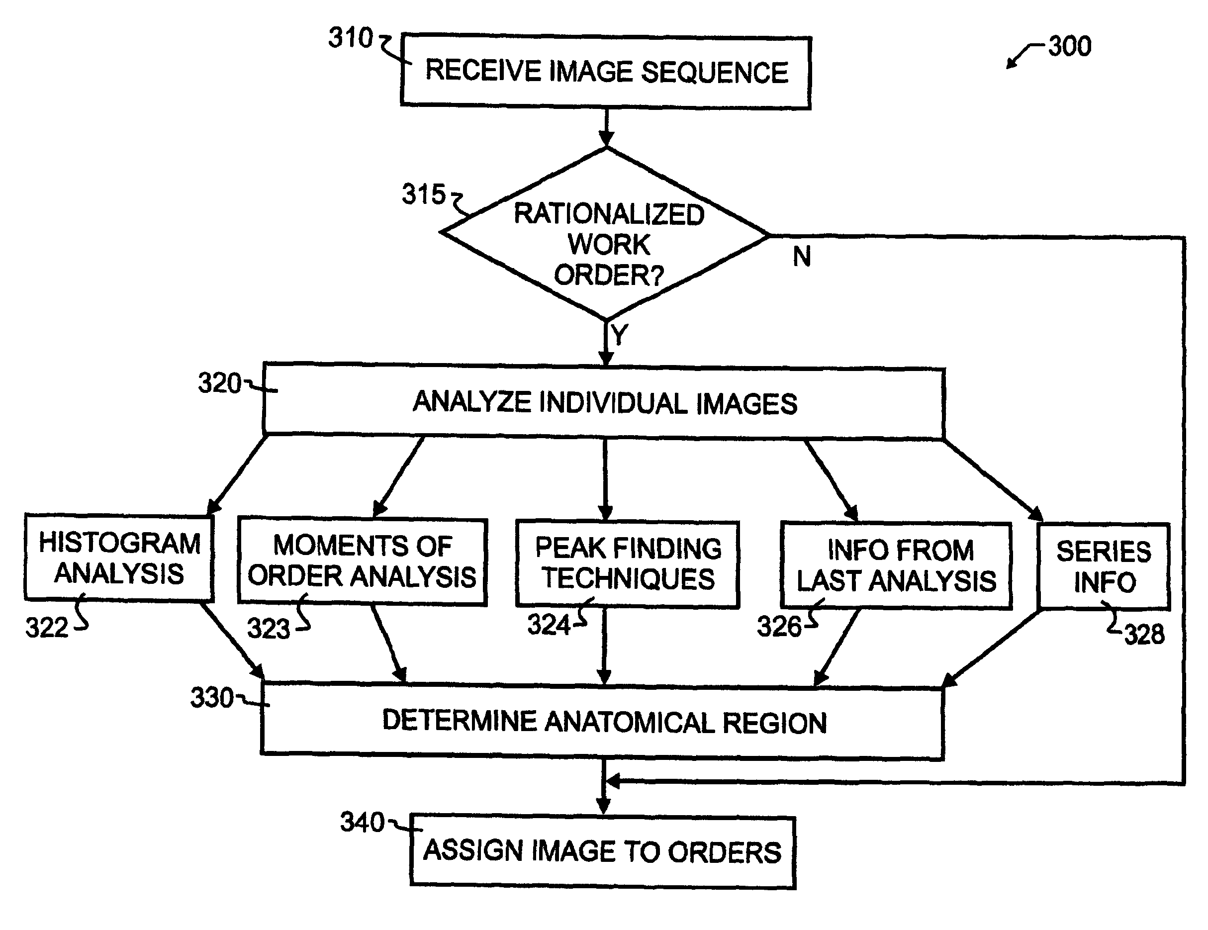
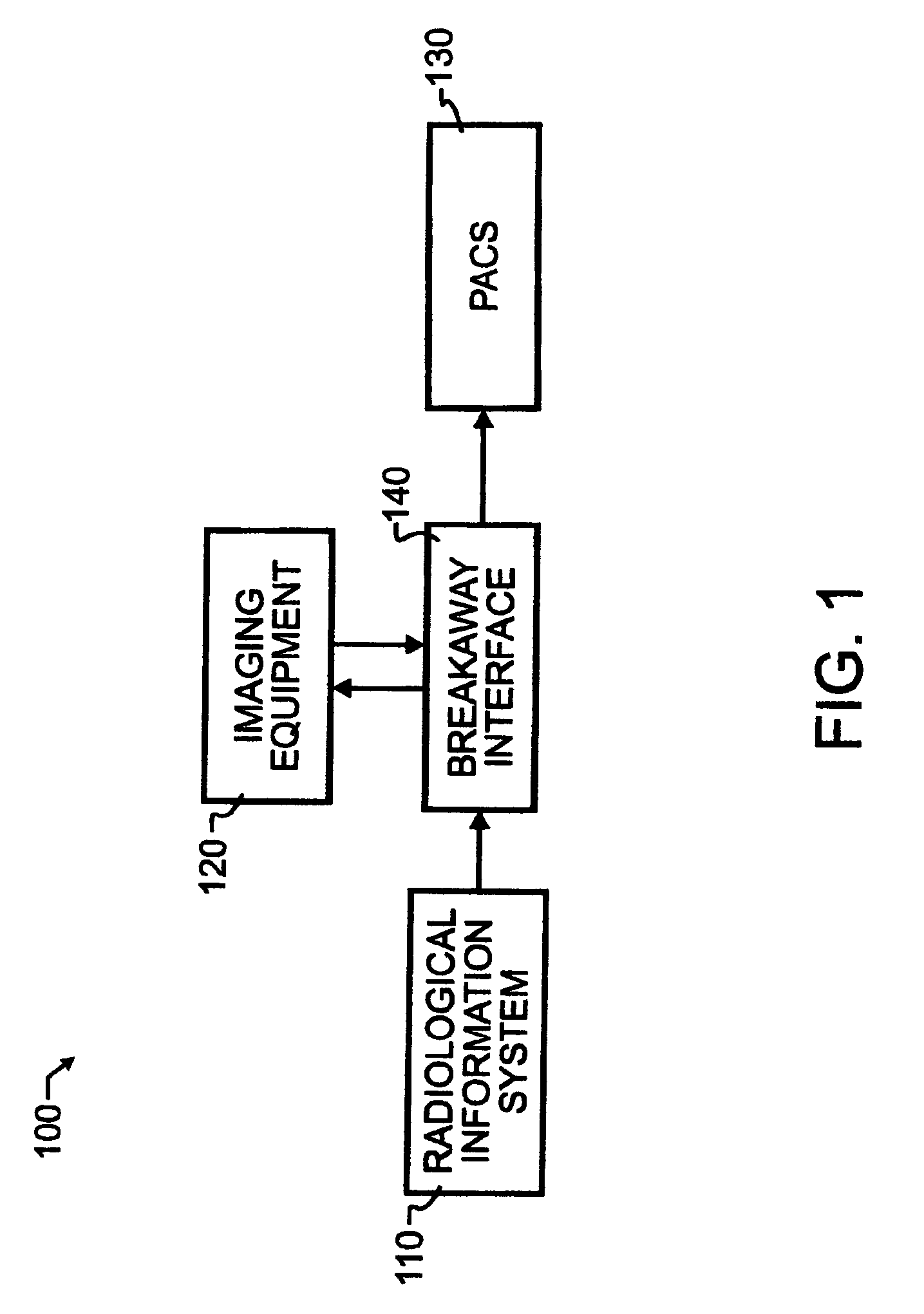
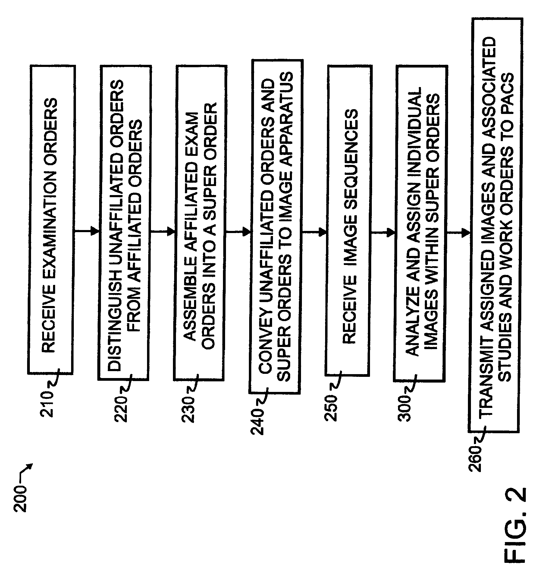
[0024]A preferred embodiment breakaway apparatus 100 and breakaway method 200 of interfacing radiological images with work orders is illustrated in FIGS. 1-3. As illustrated in FIG. 1, breakaway apparatus 100 couples a standard radiological information system 110 or other known or suitable substitute, including a PACS system, to breakaway interface 140. In the preferred embodiment, radiological information system 110 will communicate to breakaway interface 140 by transmission of communications signals using industry standard protocols such as DICOM work lists or HL-7 orders.

[0025]Most preferably, though not essential to the workings of the present invention, the various components within breakaway apparatus 100 will be compliant with the DICOM industry standards. DICOM, which stands for Digital Imaging and Communications in Medicine, is the industry standard for transfer of radiological images and other medical information between computers. Patterned after the Open System Interconn...


PUM


Abstract

A breakaway interface between radiological information systems, imaging equipment and picture archive and communications systems has automated filtering and handling of multiple study work orders or affiliated work orders, while passing single study work orders through unaltered. The work orders are processed by the breakaway interface to consolidate multiple procedure or multiple study work orders into a single super order, which is then communicated, preferably using DICOM standard protocol, to an imaging machine. The imaging machine returns a single image sequence, and the breakaway interface will then break images away from the single image sequence into a plurality of grouped image sequences. The preferred grouping is based upon anatomical regions, and separate but adjacent anatomical regions will preferably share one or more images at the boundary between the adjacent regions. The exact number of shared images may preferably be preset at the system level. A number of different techniques for analyzing the single image sequence are proposed individually or in combination, including histogram analysis, peak finding techniques, moments of order analysis, evaluating information from one or more previous analyses, and evaluating image sequence series information to distinguish discrete imaging procedures.

Description

CROSS REFERENCE TO RELATED APPLICATIONS[0001]This application claims priority to U.S. provisional application Ser. No. 60 / 437,556 filed on Dec. 31, 2002 and incorporated herein by reference in its entirety.BACKGROUND OF THE INVENTION[0002]1. Field of the Invention[0003]This invention pertains generally to computer-aided imaging systems and computer-aided radiological information systems. More specifically, the present invention incorporates image analysis and work order evaluation to ensure that a radiological information system and computer-aided imaging system produce desired and matched images and work orders in an automated and reliable manner.[0004]2. Description of the Related Art[0005]Radiological information systems provide a hospital, imaging or other medical facility appropriate computer software tools to manage information regarding radiological exams. Modern radiological imaging facilities will typically utilize a variety of highly sophisticated testing, imaging and anal...

Claims


Application Information

Patent Timeline
Patent Type & Authority Patents(United States)
IPC IPC(8): G06Q10/00G06F19/00G06Q10/10G06Q30/04G16H10/60
CPCG06F19/321G06Q10/10G06Q30/04G06Q50/22G06Q50/24G16H30/40G16H30/20
Inventor DEJARNETTE, WAYNE T.STOCKHAM, CHARLES D.WINEKE, STEVEN G.
Owner DEJARNETTE RES SYST
Why Eureka
Try Eureka