Cell analysis using laser with external cavity

a laser diode and cell technology, applied in the field of analysis and processing of biological cells, can solve the problems of large errors in detecting scattered light from an individual cell, inability to further use cells labeled with fluorescent markers, and inability to determine the properties of a single cell

Inactive Publication Date: 2010-08-03
NANYANG TECH UNIV
View PDF10 Cites 22 Cited by
  • Summary
  • Abstract
  • Description
  • Claims
  • Application Information

AI Technical Summary

Problems solved by technology

Unfortunately, all these techniques have their individual shortcomings.
First, the cell sample requires time-consuming chemical treatment and fluorescence labeling; the fluorescence labels will change the characteristics of the natural cells; moreover, those cells labeled with fluorescent markers cannot be further used, such as for transplantation.
Second, the errors of detecting scattered light from an individual cell are quite large; that is why the result from FCM is a statistic distribution and the tested sample requires certain enrichment (concentration of cells), which implies that FCM is not suitable for determining the properties of a single cell.
Third, due to the low efficiency of scattering and fluorescence from the small size of biological cells (typically less than 20 μm in diameter) and also a limited area where the fluorescence labels are excited, the signal (intensity) for each cell passing through the detection area may be weak compared to the intensity of incident laser light.
Finally, a flow cytometer is a big and expensive machine; it costs normally from $200,000 to $500,000 and needs a trained operator.
First, the fabrication for this peculiar VCSEL is very complicated and vulnerable.
Making a channel within the laser die increases the difficulty of the process.
Second, the dimension of the channel is limited by the size of VCSEL.
Moreover, this apparatus can only analyze cells with a narrow size range under a certain channel width.
Thus, big cells may be distorted in the channel and small cells might not be efficient enough in influencing the laser emission.
Third, the cells to be analyzed can not be exactly located in the middle of channel without microflow control.
Finally, it is very difficult to make the VCSEL resonant.
If the cells have a very strong absorption, the laser will not lase because of the high loss, and the cavity can not operate in resonance.

Method used


Image

  • Cell analysis using laser with external cavity
  • Cell analysis using laser with external cavity
  • Cell analysis using laser with external cavity

Examples


Embodiment Construction

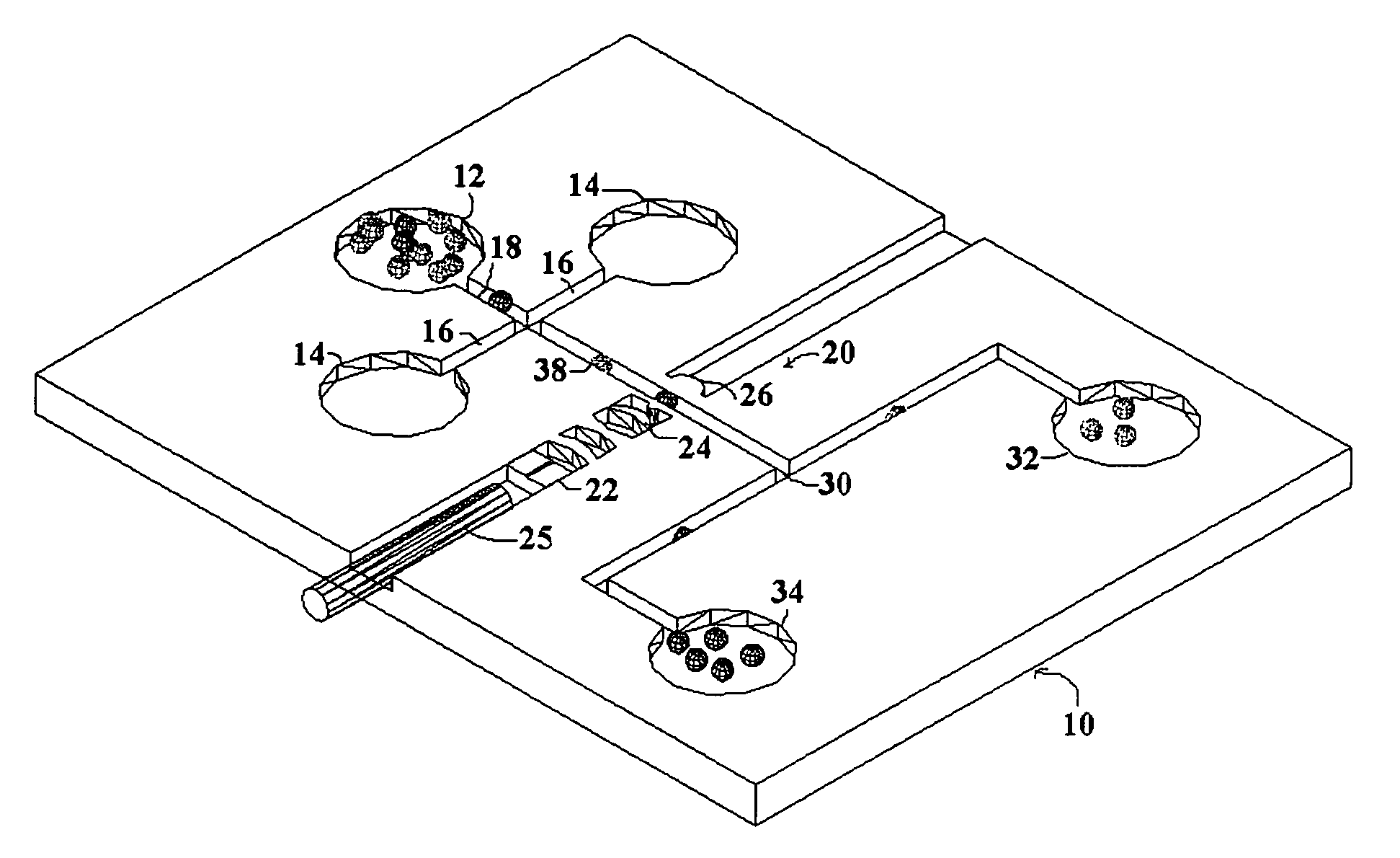
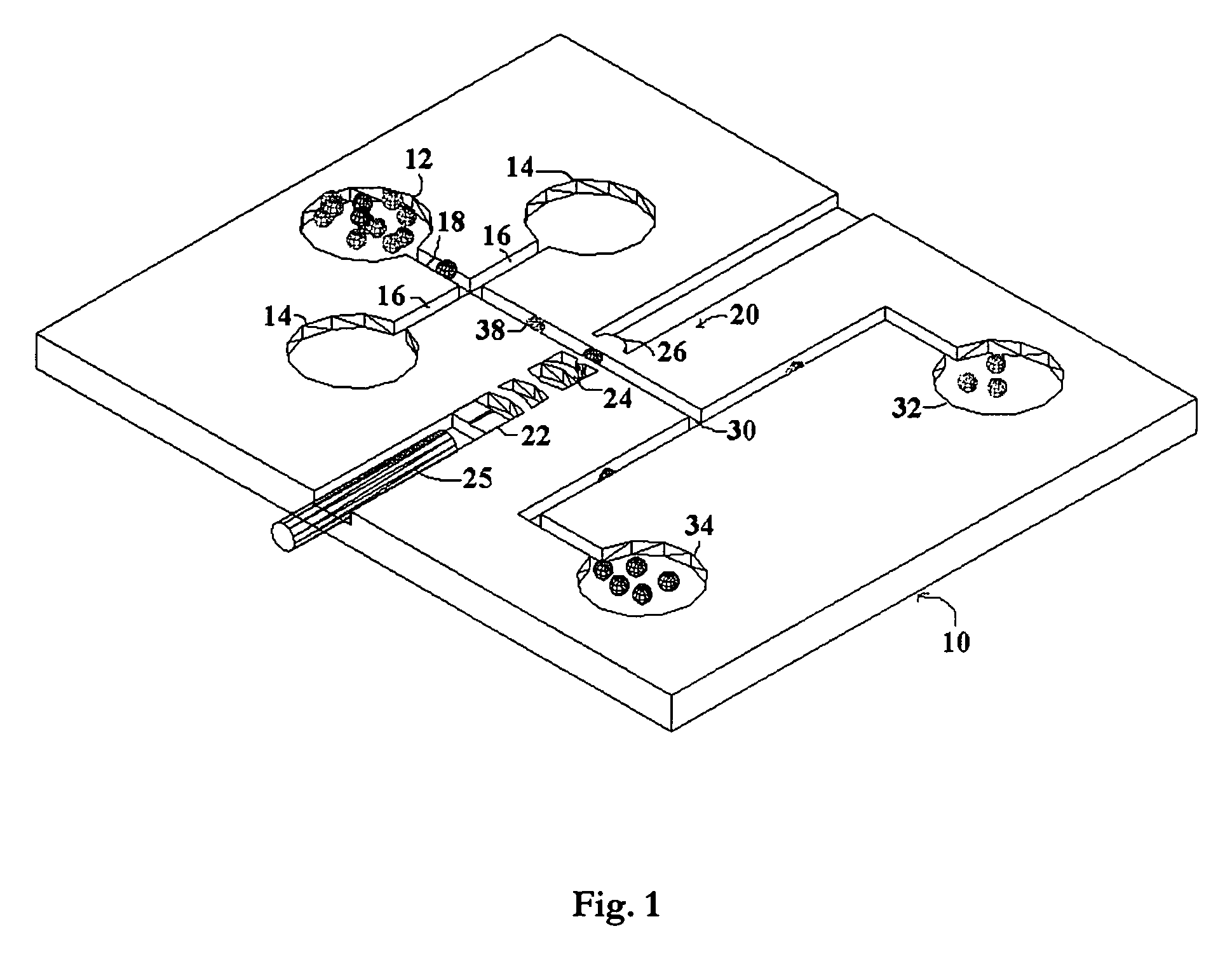
[0019]The present invention seeks to overcome the problems of the prior art by providing a system for biomedical analysis with a single operation, low costs, and high accuracy. The embodiments of the invention may independently provide one or more of the advantages set forth below.

[0020]One advantage of the present invention is that the biological cells are able to be non-invasively analyzed without any chemical treatment or fluorescent labeling, which keeps the cells intact and also enables the collected cells to be further used or processed after measurement, especially for therapeutic purposes.

[0021]Another advantage of the present invention is that the living cells are measured one by one in the analysis region in real time with high accuracy, which provides an approach for single-molecule detection (SMD) and reduces the sample consumption as well.

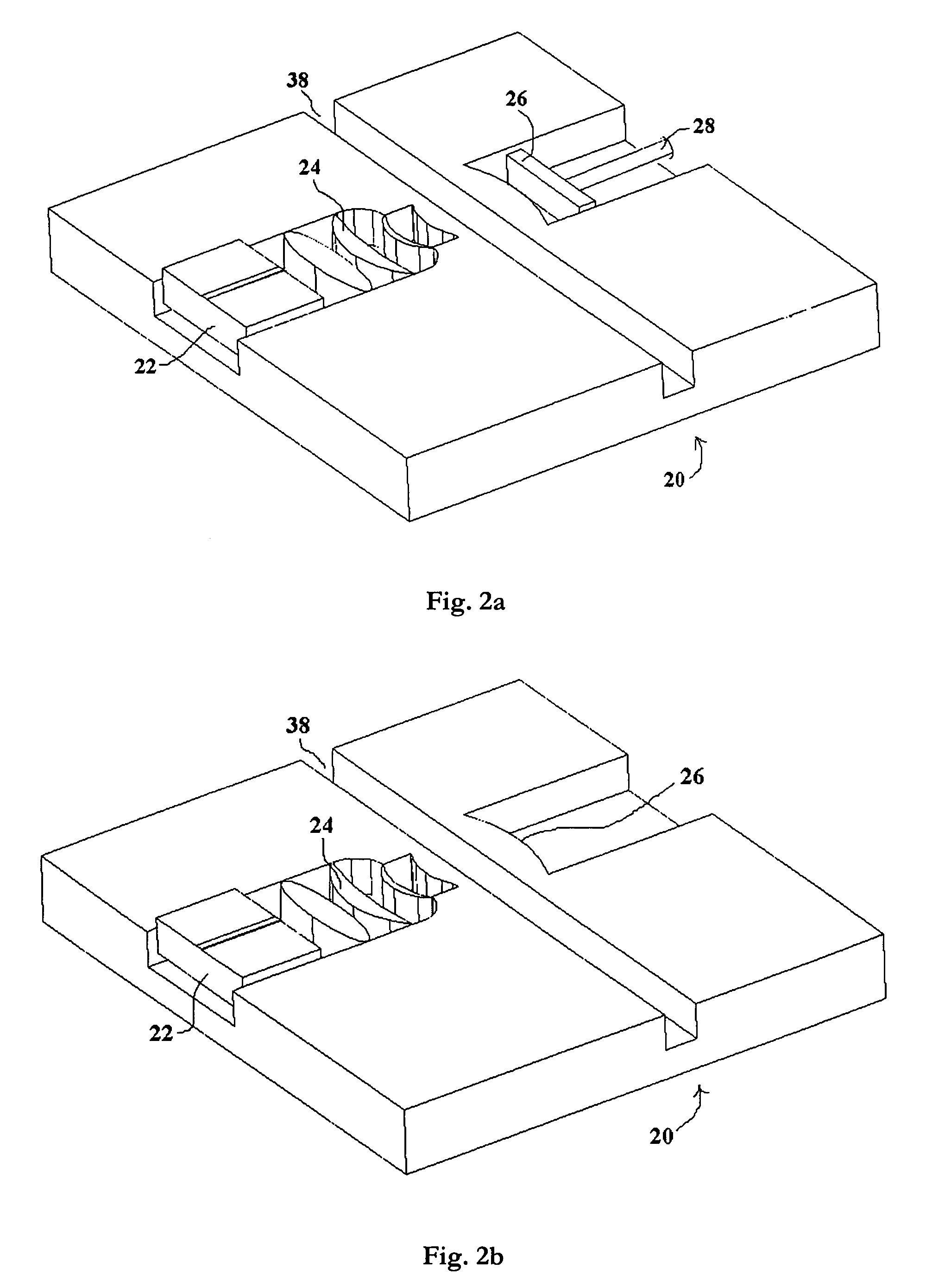
[0022]A further advantage of the present invention is that the living cells are located within an external laser cavity and analyzed ...


PUM

PropertyMeasurementUnit
reflectanceaaaaaaaaaa
diameteraaaaaaaaaa

Abstract

An apparatus and method for analyzing biological cells and other particles using an external laser cavity. Microfluidic channels contain and transport biological cells to be analyzed. A laser diode provides light for cell analysis. An external cavity is provided between one surface of the laser diode and a mirror opposite thereto. A microlens set focuses the light on only one cell as it passes through the external cavity. The presence of the cell in the external cavity gives a weak feedback toward the laser diode. The emission frequency and the output power of the laser are both functions of the length of the external cavity. Therefore, the variation of cavity length can be deduced from these parameters, where the variation is caused by changing the refractive index or size of the cell in the cavity.

Description

BACKGROUND OF THE INVENTION[0001]1. Field of the Invention[0002]This present invention relates to the analysis and processing of biological cells and other particles and more particularly to a method and device for analyzing and processing biological cells using a laser diode with an external cavity.[0003]2. Discussion of the Background[0004]In recent years, rapid in-situ monitoring of biological cells and detection of ultra-small-volume samples, and even of single cells, are in great demand (1). Those analyses and processing may include the determination of morphologic characteristics of cells and biophysical properties of cells, which is of especial importance in the fields of cytochemistry, molecular biology, genetics, immunology, and biomedicine and so forth. Two major fields are environmental monitoring and point-of-care (POC) disease diagnosis including a range of applications, from drinking water quality and food pollution to epidemics and bioterrorism.[0005]Several technique...

Claims


Application Information

Patent Timeline
Patent Type & Authority Patents(United States)
IPC IPC(8): C12M1/34G01N21/00C12M3/00
CPCG01N21/03G01N21/0303G01N21/39G01N2021/391G01N2021/399G01N2021/0346Y10S435/808
Inventor LIU, AI QUNLIANG, XIAO JUNZHANG, XU MINGSUN, YI
Owner NANYANG TECH UNIV
Why Eureka
Try Eureka