Antibodies that bind to a portion of the VWC domain of connective tissue growth factor
a technology of connective tissue growth factor and antibodies, which is applied in the direction of antibodies medical ingredients, peptide/protein ingredients, and metabolic disorders, can solve the problems of local deposition of granulation tissue, and achieve the effect of increasing the half-life of plasma
- Summary
- Abstract
- Description
- Claims
- Application Information
AI Technical Summary
Benefits of technology
Problems solved by technology
Method used
Image
Examples
example 1
Production of Recombinant Human CTGF
[0139]A recombinant human CTGF baculovirus construct was produced as described in Segarini et al. ((2001) J Biol Chem 276:40659-40667). Briefly, a CTGF cDNA comprising only the open reading frame was generated by PCR using DB60R32 (Bradham et al. (1991) J Cell Biol 114:1285-94) as template and the primers 5′-gctccgcccgcagtgggatccATGaccgccgcc-3′ and 5′-ggatccggatccTCAtgccatgtctccgta-3′, which add BamHI restriction enzyme sites to the ends of the amplified product. The native start and stop codons are indicated in capital letters.
[0140]The resulting amplified DNA fragment was digested with BamHI, purified by electrophoresis on an agarose gel, and subcloned directly into the BamHI site of the baculovirus PFASTBAC1 expression plasmid (Invitrogen Corp., Carlsbad Calif.). The sequence and orientation of the expression cassette was verified by DNA sequencing. The resulting CTGF expression cassette was then transferred to bacmid DNA by site-specific recom...
example 2
CTGF N-Terminal and C-Terminal Fragment Production
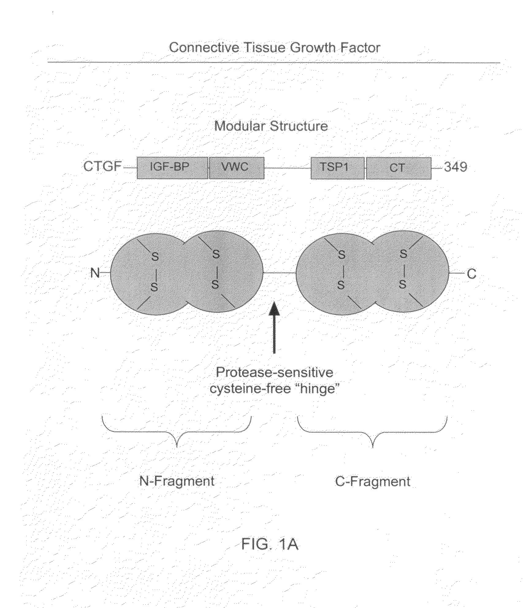
[0145]N-terminal fragments and C-terminal fragments of CTGF were prepared as follows. Recombinant human CTGF, prepared and purified as described above, was digested at room temperature for 6 hours by treatment with chymotrypsin beads (Sigma Chemical Co., St. Louis, Mo.) at 1.5 mg of CTGF per unit of chymotrypsin. The mixture was centrifuged, the chymotrypsin beads were discarded, and the supernatant, containing enyzmatically-cleaved rhCTGF, was diluted 1:5 with 50 mM Tris, pH 7.5. The diluted supernatant was applied to a Hi-Trap heparin column. The flow-through, containing N-terminal fragments of CTGF, was collected. The heparin column was washed with 350 mM NaCl, and bound C-terminal fragments of CTGF were eluted with a linear gradient of 350 mM to 1200 mM NaCl, as described above. The fractions were analyzed by SDS-PAGE, and fractions containing C-terminal fragments of CTGF were pooled.
[0146]The heparin column flow-through, which c...
example 3
Production of Human Anti-CTGF Monoclonal Antibodies
[0147]Fully human monoclonal antibodies to human CTGF were prepared using HUMAB mouse strains HCo7, HCo12 and HCo7+HCo12 (Medarex, Inc., Princeton N.J.). Mice were immunized by up to 10 intraperitoneal (IP) or subcutaneous (Sc) injections of 25-50 mg recombinant human CTGF in complete Freund's ajuvant over a 2-4 weeks period. The immune response was monitored by retroorbital bleeds. Plasma was screened by ELISA (as described below), and mice with sufficient titers of anti-CTGF immunogolobulin were used for fusions. Mice were boosted intravenously with antigen 3 and 2 days before sacrifice and removal of the spleen.
[0148]Single cell suspensions of splenic lymphocytes from immunized mice were fused to one-fourth the number of P3×63-Ag8.653 nonsecreting mouse myeloma cells (American Type Culture Collection (ATCC), Manassas Va.) with 50% PEG (Sigma, St. Louis Mo.). Cells were plated at approximately 1×10⁵ cells / well in flat bottom micro...
PUM
 Abstract
Description
Claims
Application Information
 - R&D
- Intellectual Property
- Life Sciences
- Materials
- Tech Scout
- Unparalleled Data Quality
- Higher Quality Content
- 60% Fewer Hallucinations
Browse by: Latest US Patents, China's latest patents, Technical Efficacy Thesaurus, Application Domain, Technology Topic, Popular Technical Reports.
© 2025 PatSnap. All rights reserved.Legal|Privacy policy|Modern Slavery Act Transparency Statement|Sitemap|About US| Contact US: help@patsnap.com


