Methods of treating a trauma or disorder of the knee joint by local administration and sustained-delivery of a biological agent

a biological agent and knee joint technology, applied in the field of knee joint trauma or disorder treatment, can solve the problems of limited quantity of agents, insufficient methods to serve the needs of patients, and degradation of surrounding tissues and associated chronic pain

Inactive Publication Date: 2011-03-22
WARSAW ORTHOPEDIC INC
View PDF30 Cites 18 Cited by
  • Summary
  • Abstract
  • Description
  • Claims
  • Application Information

AI Technical Summary

Benefits of technology

[0006]The present invention relates to a method of providing a subject with sustained-release of a biological agent within a joint such as, but not limited to, the knee joint. This method involves minimally invasively or surgically attaching a pharmaceutical depot implant containing the biological agent to a ligament, tendon, muscle or other structure within the joint so as to provide sustained-release of the agent to treat inflammation, a trauma, and / or disorder in the joint, while allowing for normal joint articulation.
[0009]The present invention also relates to methods wherein multiple pharmaceutical depot implants may be minimally invasively or surgically secured to both the anterior cruciate ligament and the posterior cruciate ligament, thus promoting the prolonged prophylactic or therapeutic effects contemplated herein.
[0012]The present invention also relates to methods wherein multiple pharmaceutical depot implants may be minimally invasively or surgically secured to the anterior cruciate ligament, the posterior cruciate ligament, the patellar ligament, the medial collateral ligament, and / or the lateral collateral ligament, or any combination thereof, so as to afford maximum therapeutic or prophylactic effect from the sustained-release of a biological agent from the respective depot implant(s).
[0013]In one embodiment, the injury or disorder is associated with an intraarticular ligament (e.g., the ACL and PCL). In another embodiment the trauma or disorder effects other ligaments of the knee joint (including but not limited to the lateral or medial collateral ligaments, as well as the patellar ligament). In other embodiments, the injuries or disorders relate to problems associated with the articular cartilage, such as osteoarthritis, chondromalacia, and rheumatoid arthritis. Another embodiment relates to treating meniscal injuries, such as meniscal tears. Additional embodiments include, but are not limited to, chondral fractures, traumas or injuries associated with the patella, just to name a few. Thus, the methods of the present invention may be utilized to deliver a biological agent to the knee joint area in a prolonged, sustained time frame and provide therapeutic or prophylactic treatment of any trauma or disorder of the knee while allowing for normal articulation of the knee joint.
[0019]Another object of the present invention is to provide for sustained-delivery of a biological agent from an implanted drug delivery device within a synovial joint such that the drug treats joint inflammation, trauma and / or a disorder. Accordingly, the use of the device obviates the need for regular dosing by the patient and increases patient compliance with either a prescribed therapeutic regimen or a prophylactic regimen prescribed prior to the onset of symptoms.
[0020]Another object of the present invention is to provide for sustained-delivery of a biological agent from an implanted drug delivery device within the knee joint such that the drug treats a knee trauma or knee disorder. Accordingly, the use of the device obviates the need for regular dosing by the patient and increases patient compliance with either a prescribed therapeutic regimen or a prophylactic regimen prescribed prior to the onset of symptoms.

Problems solved by technology

Left unchecked, this can lead to degradation of surrounding tissues and associated chronic pain.
TNF-a inhibitors currently in use are generally administered systemically via intravenous infusion and subcutaneous injection, but there are side effects of anti-TNF therapies associated with the higher doses and systemic administration that are common with these therapies.
Unfortunately, it provides a limited quantity of agent that must move through the tissue to the target site.
Such methods are inadequate to serve the needs of patients.

Method used


Image

  • Methods of treating a trauma or disorder of the knee joint by local administration and sustained-delivery of a biological agent
  • Methods of treating a trauma or disorder of the knee joint by local administration and sustained-delivery of a biological agent
  • Methods of treating a trauma or disorder of the knee joint by local administration and sustained-delivery of a biological agent

Examples


first embodiment

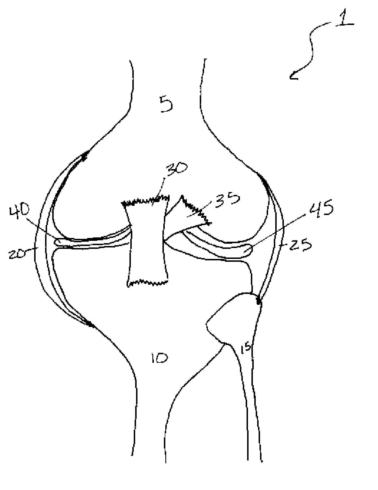
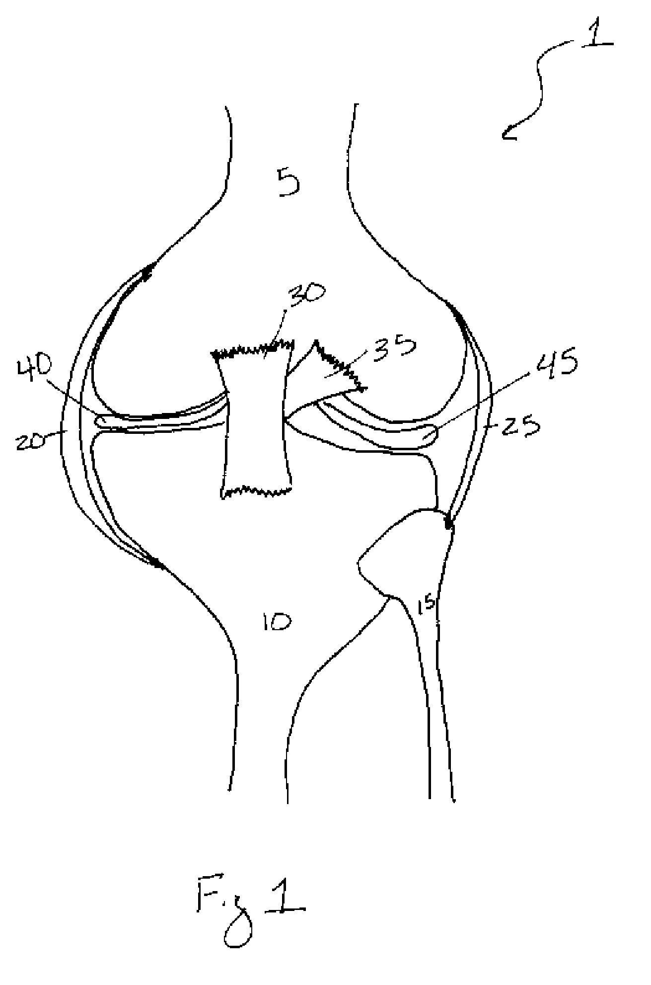
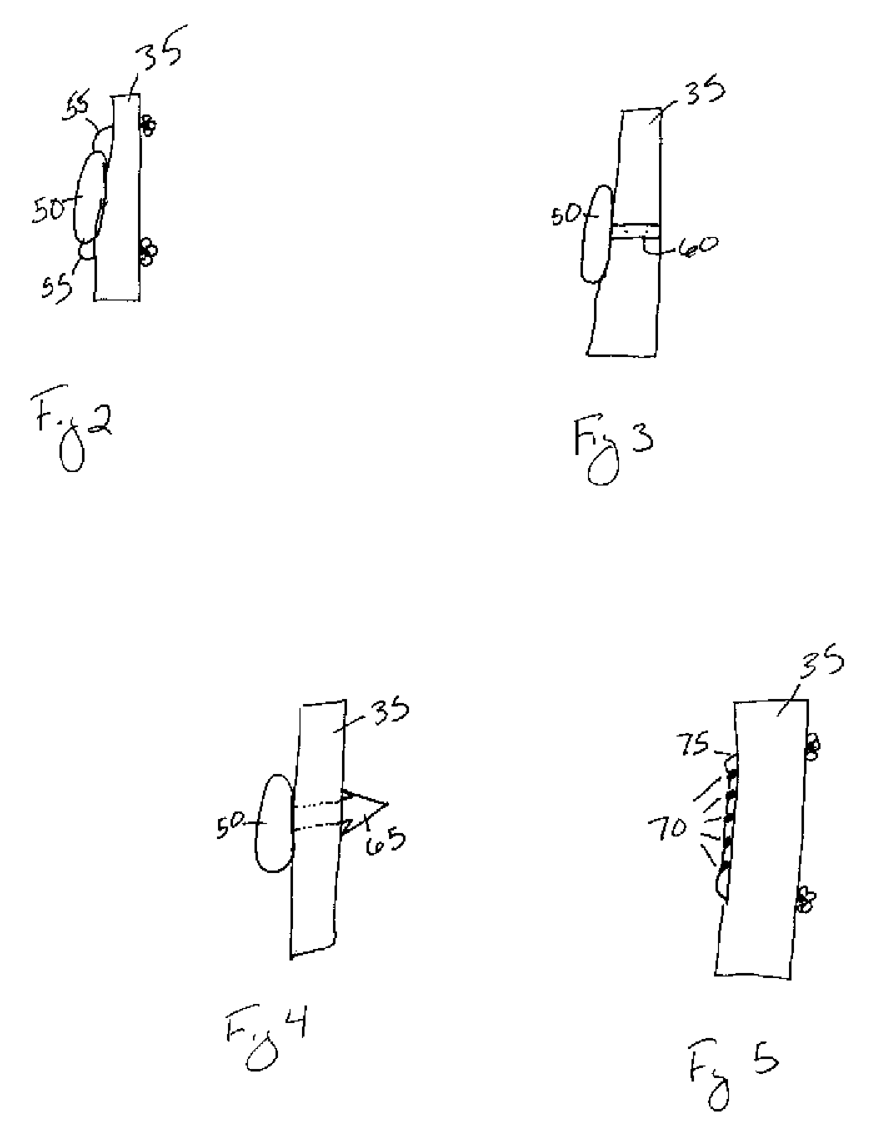
[0070]Referring to FIG. 2, in a first embodiment the depot may be comprised of a monolithic or coaxially extruded rod 50. At least one suture 55 may extend from either or both of the ends of the rod 50. The sutures 55 may be comprised of any natural or synthetic, biocompatible and biodegradable polymer, or of any material understood in the art to secure a depot to soft tissue. The suture 55 may also extend from the center of the depot or any location on the depot understood to secure the depot to the soft tissue. In operation, FIGS. 2 and 8 illustrate the product of one method of securing the depot to the ACL 35 by tying the depot to the ACL 35 such that the depot is positioned in the center of the ACL 35 to ensure uniform distribution of the biological agent within the synovial joint. The suture 55 may secure the depot to the ACL 35 by threading the suture(s) through microincisions in the ligament, by tying the suture(s) substantially around the ligament, or any similar method unde...

second embodiment

[0071]Referring to FIG. 3, in a second embodiment the depot may be comprised of a rod 50 with at least one strap or clasp 60 extending from the rod 50. The strap / clasp 60 may be comprised of any natural or synthetic, biocompatible and biodegradable polymer, or of any material understood in the art to secure a depot to soft tissue using a strap / clasp 60. The strap / clasp 60 may extend from the center of the rod 50 or from any location on the rod 50 understood to secure the rod 50 to the soft tissue. In operation, the strap / clasp 60 may be secured to a ligament, such as the ACL 35, within the joint capsule. FIGS. 3 and 8 illustrate the product of one method of securing the depot to the ACL 35 such that the strap / clasp 60 substantially surrounds the ACL 35. Moreover, the depot is positioned in the center of the ACL 35 to ensure uniform distribution of the biological agent within the synovial joint. The rod 50 may also be secured to the ACL 35 distal to the PCL 30, or in any location, su...

third embodiment

[0072]Referring to FIG. 4, in a third embodiment the depot may be comprised of a rod 50 with at least one barb 65 extending from the rod 50. The barb 65 may be comprised of any natural or synthetic, biocompatible and biodegradable polymer, or of any material understood in the art to secure a depot to soft tissue using at least one barb 65. The barb 65 may extend from the center of the rod 50 or from any location on the rod 50 understood to secure the rod 50 to the soft tissue. In operation, the barb 65 is secured to a ligament, such as the ACL 35, within the joint capsule. FIG. 4 illustrates one method of securing the depot to the ACL 35 wherein the barb 65 is threaded through a microincision in the ACL 35. The depot is positioned in the center of the ACL 35 to ensure uniform distribution of the biological agent within the synovial joint. The rod 50 may also be secured to the ACL 35 distal to the PCL 30 or at any location of the ACL 35 so long as the depot does not interfere with th...


PUM

PropertyMeasurementUnit
diameteraaaaaaaaaa
diameteraaaaaaaaaa
molecular massaaaaaaaaaa

Abstract

Methods and apparatus of providing a subject with post-operative, sustained-release of a biological agent within a synovial joint is disclosed. These methods involve securing a depot containing the biological agent to a ligament, tendon, muscle within the joint to provide sustained-release of the agent while allowing for normal joint articulation. This methodology may be utilized to provide for sustained-release of a biological agent useful in treating various traumas and disorders of the joint. Such biological agents include antagonists of inflammation-related proteins, such as TNF-α, IL-1β, IL-6, IL-8, NF-κB, High Mobility Group Box 1 (HMG-B1), IL-2, IL-15 and steroidal and non-steroidal anti-inflammatories. Other biological agents include anti-inflammatory cytokines such as IL-10, IL-4, IL-13, and TGF-β. The biological agents also include osteogenic and cartilage producing growth factors such as, but not limited to, BMP-2, BMP-4, BMP-6, BMP-7, BMP-8, and MIA CD-RAP. Finally, the biological agents include siRNA and / or therapeutic antibodies.

Description

FIELD OF THE INVENTION[0001]The present invention relates to methods of treating a trauma or disorder of a joint comprising delivery of a biological agent to the joint as a depot implant while also providing for normal articulation of the joint. Embodiments of the methods of the present invention include minimally invasive or surgical attachment of a pharmaceutical depot implant containing a biological agent to a ligament of a joint such as, but not limited to, a knee joint. The methods of the present invention may be utilized to treat pain and inflammation associated with the cartilage, subchondral bone, ligament, tendon and / or muscle of the joint. These methods are also contemplated for the prophylactic or therapeutic treatment of various traumas or disorders of the knee joint, including but not limited to primary repair or reconstruction of the anterior or posterior cruciate ligament, the medial or lateral collateral ligament, the patellar ligament, or disorders such as osteoarth...

Claims


Application Information

Patent Timeline
Patent Type & Authority Patents(United States)
IPC IPC(8): A61F2/02A61K38/16A61K38/20A61M31/00A61P21/00
CPCA61K38/1841A61K38/1875A61K38/2026A61K38/2066A61M31/002A61P21/00
Inventor MCKAY, WILLIAM F.
Owner WARSAW ORTHOPEDIC INC
Why Eureka
Try Eureka