Phosphatase inhibitor protein-1 as a regulator of cardiac function

a phosphatase inhibitor and protein technology, applied in the direction of cardiovascular disorders, peptides, drug compositions, etc., can solve the problems of heart failure to the rest of the body, inability to detect any inhibition of pp-1 activity, and insufficient human resources, so as to improve the end-systolic pressure dimension relationship, accelerate calcium signal decay, and reduce the time constant for relaxation

Active Publication Date: 2011-08-02
UNIVERSITY OF CINCINNATI
View PDF3 Cites 8 Cited by
  • Summary
  • Abstract
  • Description
  • Claims
  • Application Information

AI Technical Summary

Benefits of technology

[0021]In another aspect, the invention provides a method of decreasing cardiac contractility in a subject, the method comprising introducing, into heart cells of the subject, an effective amount of an isolated nucleic acid molecule that encodes a constitutively unphosphorylated phosphatase inhibitor-1 protein comprising the amino acid sequence of SEQ ID NO: 5 or SEQ ID NO: 6, or a constitutively unphosphorylated fragment thereof, thereby decreasing cardiac contractility in the subject.
[0022]In another aspect, the invention provides a method of decreasing cardiac contractility in a subject, the method comprising administering an effective amount of an isolated polypeptide comprising the amino acid sequence of SEQ ID NO: 5 or SEQ ID NO: 6, or a constitutively unphosphorylated fragment thereof, thereby decreasing cardiac contractility in the subject.
[0028]In another embodiment of a method according to the invention, the nucleic acid molecule is introduced in an amount effective to result in a condition selected from the group consisting of myocyte shortening, lowering of the time constant for relaxation, and accelerating calcium signal decay, and combinations thereof. In yet another embodiment, the nucleic acid molecule is introduced in an amount effective to improve the end-systolic pressure dimension relationship.

Problems solved by technology

However, in vitro testing conditions failed to reveal any inhibition of PP-1 activity.
Heart failure is almost always a chronic, long-term condition, and consumes an inordinate amount of medical intervention and human resource dollars.
In particular, the consequences of heart failure to the rest of the body organs can be devastating both in terms of the overall reduction in productive life of the patient, and the expense of treatment.
Many organs and organ systems begin to suffer cumulative damage from lack of oxygen and nutrients.

Method used


Image

  • Phosphatase inhibitor protein-1 as a regulator of cardiac function
  • Phosphatase inhibitor protein-1 as a regulator of cardiac function
  • Phosphatase inhibitor protein-1 as a regulator of cardiac function

Examples


example 1

Phosphorylation of I-1 and I-1 Mutants by PKC-α

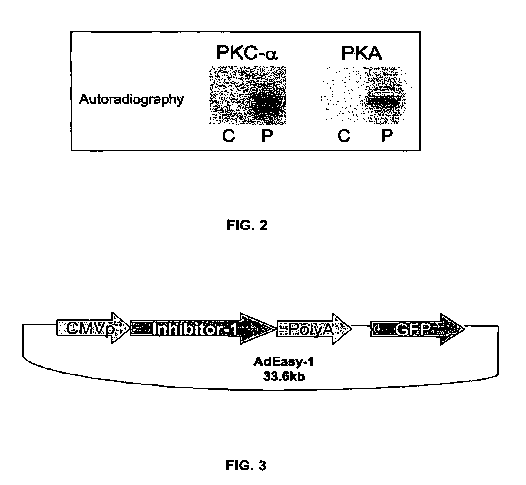
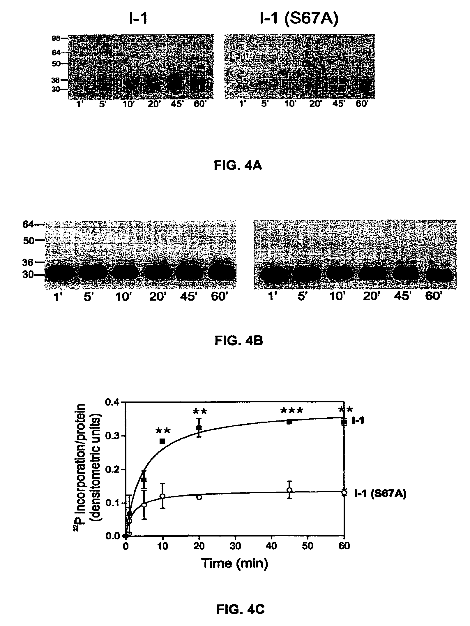
[0177]As noted, previous work showed that PKC-α phosphorylates inhibitor-1 (I-1) at Ser-67. To examine whether additional PKC-α phosphorylation sites may exist on I-1, an I-1 mutant in which Ser-67 was substituted with alanine was phosphorylated in vitro by PKC-α as described above. FIG. 4A shows that although PKC-α phosphorylation of the mutant I-1 (S67A) is greatly decreased in comparison to wild type I-1, it is not completely abolished. Densitometric analysis of 32P-incorporated per protein revealed that at steady-sate (20-45 min), I-1 (S67A) incorporated 40±8.6% of the radioactivity levels present in wild types (100%) (FIG. 4C). In some of the experiments, a single additional band appeared at 78 Kda, which correspond to PKC-α autophosphorylation. No other radioactive bands were detected in any of the experiments. A specific antibody (AC1, 1:1000) recognized the phospho-bands as inhibitor-1 (FIG. 4B). Thus, these results indicate tha...

example 2

Phosphorylation Site Determination

[0178]To determine the location of the additional PKC-α site (s) on I-1, recombinant purified I-1 was purified and subjected to in vitro phosphorylation by PKC-α in the presence of [γ-32P] ATP. The 32P-labeled I-1 was purified by SDS-PAGE, and digested with trypsin. After purification of the tryptic peptides by reverse-phase HPLC, two peaks, 50 and 51 (FIG. 5A), contained 62% of the radioactivity eluting from the Vydac column. Both fractions were subjected to MALDI-TOF mass spectrometry analysis. Mass matching analysis from fraction 51 yielded three potential phosphorylated peptides. The detected masses (predicted mass plus phosphate group) were 1226.44, 1494.45, and 1572.68 Da, which corresponded to each of the following I-1 sequences: 62STLAMSPRQR71 (SEQ ID NO: 31), 72KKMTRITPTMK82 (SEQ ID NO: 23), and 135KTAECIPKTHER146 (SEQ ID NO: 32) (data not shown). In parallel, analysis of peak 50 detected a peptide of mass 1366.46 Da, which corresponded to ...

example 3

Analysis of PKC-α Phosphorylation of I-1 by Two-Dimensional Electrophoresis

[0182]To further corroborate the autoradiography results, 2-D gel electrophoresis analysis was used to detect possible mobility changes in I-1 due to PKC-α phosphorylation. 2-D gel electrophoresis separates proteins based on both their isoelectric point (pI) and molecular weight. Phosphorylation causes the pI value of a protein to become more acidic, but has a negligible effect on its molecular weight. Analysis of the non-phosphorylated I-1 gel image indicated that the protein migrates in a 2-D gel as a single spot at a pI of 5.1 and molecular weight of ˜30 kD (FIG. 7A). In the PKC-α phosphorylated sample, three spots were visible, with pIs of (from right to left): 5.1, 4.9 and 4.7, corresponding to non-phosphorylated, singly phosphorylated, and doubly phosphorylated protein, respectively (FIG. 7B). A higher concentration of phosphorylated I-1 (35 ug) subjected to 2D gel electrophoresis did not show any addit...


PUM


Abstract

The present invention relates to novel nucleic acids which encode novel mutant forms of Inhibitor Protein-1 (1-1). In particular, the 1-1 mutant forms comprise altered phosphorylation sites of PKC-α. In addition, the present invention relates to methods of regulating cardiac contractility and function, and for treatment of cardio myopathy and heart failure, which employ the novel nucleic acids and polypeptides. Vectors comprising the novel nucleic acids, Antibodies to the novel proteins, and diagnostic and screening methods associated therewith, are also provided.

Description

RELATED APPLICATIONS / PATENTS & INCORPORATION BY REFERENCE[0001]This application is the U.S. national phase, pursuant to 35 U.S.C. §371, of PCT international application Ser. No. PCT / US2007 / 003470, filed Feb. 9, 2007, designating the United States and published in English on Sep. 7, 2007 as publication no. WO 2007 / 100465 A2, which claims priority to U.S. Provisional Application Ser. No. 60 / 772,327, filed Feb. 10, 2006. The entire contents of the aforementioned patent applications are incorporated herein by this reference.STATEMENT OF GOVERNMENT SUPPORT[0002]This invention was made with Government support under Grant Nos. DK036569, HL007382, HL026057, HL052318, HL057263, HL057623, HL064018, and HL077101 awarded by the National Institutes of Health. The Government has certain rights in this invention.SEQUENCE LISTING[0003]The instant application contains a Sequence Listing which has been submitted in ASCII format via EFS-Web and is hereby incorporated by reference in its entirety. Said...

Claims


Application Information

Patent Timeline
Patent Type & Authority Patents(United States)
IPC IPC(8): C07H21/02C12N15/12
CPCC07K14/4703A61K38/00A61P9/00A61P9/04
Inventor KRANIAS, EVANGELIARODRIGUEZ, PATRICIAMITTON, BRYAN
Owner UNIVERSITY OF CINCINNATI
Why Eureka
Try Eureka