Ultrasonic knife

a cutting device and ultrasonic technology, applied in the field of ultrasonic cutting devices, to achieve the effects of promoting the capability of tipping, promoting safety, and facilitating the sleeve cover

Inactive Publication Date: 2011-09-13
ALCON INC
View PDF60 Cites 64 Cited by
  • Summary
  • Abstract
  • Description
  • Claims
  • Application Information

AI Technical Summary

Benefits of technology

[0016]The subject matter of the present invention includes a cutting device as in an ultrasonic cutting device designed to cut a cataract in sizable and manipulatable fragments with little difficulty, unlike the standard phacoemulsification cylindrical needles which are not well suited to easily divide the cataract in that manner. A preferred embodiment features an ultrasonic cutting device comprising a shaft, which may be hollow or solid, and an opposing side walled “flattened” tip disposed at a distal end of the shaft. This tip vibrates at ultrasonic frequency, and is suitable for replacing the mechanical energy used by choppers and pre-choppers thus providing for efficient usage of ultrasonic energy. The ultrasonic knife of the subject matter of the present invention makes it possible to easily and efficiently divide the cataract in as many fragments as the surgeon deems desirable (e.g., 4 to 12). The cutting device of the present invention provides a tip design that easily and efficiently cuts through a wide assortment of cataract types (e.g., lower energy usage and associated lowering of heat generation with less trauma potential). For example, the cutting device of the present invention works in softer cataracts like “a hot knife cutting through butter”, and in harder cataracts (including brunescent and black cataracts) like “a hot knife cutting through a chocolate bar”.
[0017]The new ophthalmologic cutting device of the present invention provides a tool which permits a surgeon to improve on the phacoemulsification technique, making it easier to divide the cataract into several manipulatable fragments which can then be, for example, more easily emulsified with a standard phacoemulsification needle, diminishing the amount of ultrasonic energy applied during cataract extraction, and thus reducing the risk of damage to the cornea. In addition, the produced fragments can also be more easily liquefied with technologies that employ fluid pulses, making the present cutting device even more applicable to harder cataracts.
[0018]The cutting device of the present invention thus provides an “ultrachopper” that makes for easier performance of a phacoemulsification technique; as in a phacoemulsification technique that includes a breaking apart of the cataractous crystalline lens into more manageable pieces via the ultrachopper to facilitate completion of the phacoemulsification process as by applying a vibrating aspirating needle on the more manageable pieces created with the ultrachopper. Another alternative to finish the cataract extraction following the fracture with the ultrachopper is using a liquid application tool (e.g., a heated and pulsing liquefaction based further breakdown tool) to liquefy and aspirate the fragments. This procedure provides a hybrid, phacoemulsification / liquefaction procedure (which is categorized under the present invention as a phacoemulsification procedure and phacoemulsification tooling in general). Thus, with the ultrachopper, surgeons who are not highly experienced may learn these techniques in less time and with fewer complications, and experienced surgeons may improve even more their outcomes.

Problems solved by technology

The needle, however, is not designed for cutting the nucleus of the cataract or dividing it in several sizable fragments.
The needle involved in many prior art techniques has a cylindrical shape with a diameter between 1.0 to 1.5 mm, and thus is not well suited for cutting or separating the cataract in sizable fragments, which is the preferred approach taken in modern phacoemulsification techniques.
This has been shown to be able to produce damage to the eye, if applied for a long enough period of time.
Moreover, even with the application of high ultrasonic energy levels, the dividing of a very advanced cataract still may not be possible using the method of sculpting grooves, as these cylindrical hollow needles are not really adequate to divide the cataract in sizable fragments by themselves.
However, phaco chop techniques can be more technically difficult, leading to a greater likelihood of complications, and, moreover, dividing hard nucleus (brunescent and black) cataracts with pre-chopping techniques may be very difficult to master by surgeons.
As will become more apparent below (e.g., see the Summary of Invention discussion below) the cutting phaco tip, has a design that is not efficient in many respects in the cutting and fragmentizing of the wide range of cataract types faced.
Due to the limitations as to current phacoemulsification surgical instruments available, ophthalmologists around the world rely on basically the same phacoemulsification surgical technique, with some minor variations.
1. The providing of an access incision which is typically corneal or scleral with a size often varying between 1 and 3.2 mm. The incision may be located superiorly (12 or 11 o'clock positions), or completely temporal. Incision architecture may vary. There may be utilized single plane, two plane or three plane incisions, depending on the surgeon's preference. All of these incisions are intended to be watertight and do not require sutures to close.
2. Capsulorhexis is the surgical step where a central circular portion of the anterior capsule is removed and it leaves the anterior cortex of the cataract exposed. In other words capsulotomy involves creating a continuous tear of the anterior wall to produce a smooth-edged round opening. The continuous tear capsulotomy is known as “capsulorhexis”. Such a capsulotomy facilitates removal of the old lens and also facilitates in-the-bag implantation of an intraocular lens. There are several modes of capsulorhexis forceps and as some examples reference is made to U.S. Publication 2004 / 0116950 A1 to Elibschitz—Tsimhoni and U.S. Publication 2005 / 0228419 to El-Mansoury for a discussion of capsulotomy techniques and instruments.
3. Hydrodisection is a maneuver by which, using balanced saline solution injection, the cortex of the cataract is separated from the capsules, so that the cataract can rotate inside the bag.
4. Hydrodelineation is a maneuver by which, using balanced saline solution, the nucleus of the cataract is separated from the most peripheral portion of the cataract that is epinucleus and cortex, so that the nucleus can rotate freely.
5. Nuclear pre-fracture is a more recent cataract surgical technique wherein, before trying to emulsify a cataract, the nucleus is divided into sizable fragments by one of several methods, such as those described above, in order to facilitate its extraction. As noted above, since the phacoemulsification cylindrical needles can emulsify and aspirate just one small portion of the cataract at a time, and not big portions of the cataract, the pre-fracture technique facilitates the complete removal of the cataract material as with a subsequently applied cylindrical phacoemulsification needle. One pre-fracture method involves sculpting deep grooves inside the cataract with the phaco needle to provide for the fragmentation using the needle, and a second instrument to push the fragments apart. The downside of this technique is that it requires more ultrasonic energy, especially in hard and black cataracts, and this high energy level requirement can cause damage to the cornea. Moreover, using this method sometimes it can be impossible to divide certain very hard cataracts (like black cataracts), and it may be necessary to widen the surgical incision to extract it in one piece and then suture it, which is undesirable from a post treatment healing standpoint.
6. Other alternative techniques to divide the cataract before emulsifying it, is using the choppers and the prechoppers (like the above described Nagahara's chopper and Akahoshi's prechopper), which use mechanical energy. They have the drawback of being difficult to learn, taking a long time for the surgeon to master these techniques. Moreover they have the risk of rupture of the anterior capsule edge (in the case of choppers) or zonular stress (in the case of prechoppers). In very hard nucleus (brunescent and black cataracts), these techniques are not easy to perform, even for the experienced surgeon, since the fragments do not separate completely, but their fibers are stretched without breaking.

Method used


Image

  • Ultrasonic knife
  • Ultrasonic knife
  • Ultrasonic knife

Examples


embodiment 46

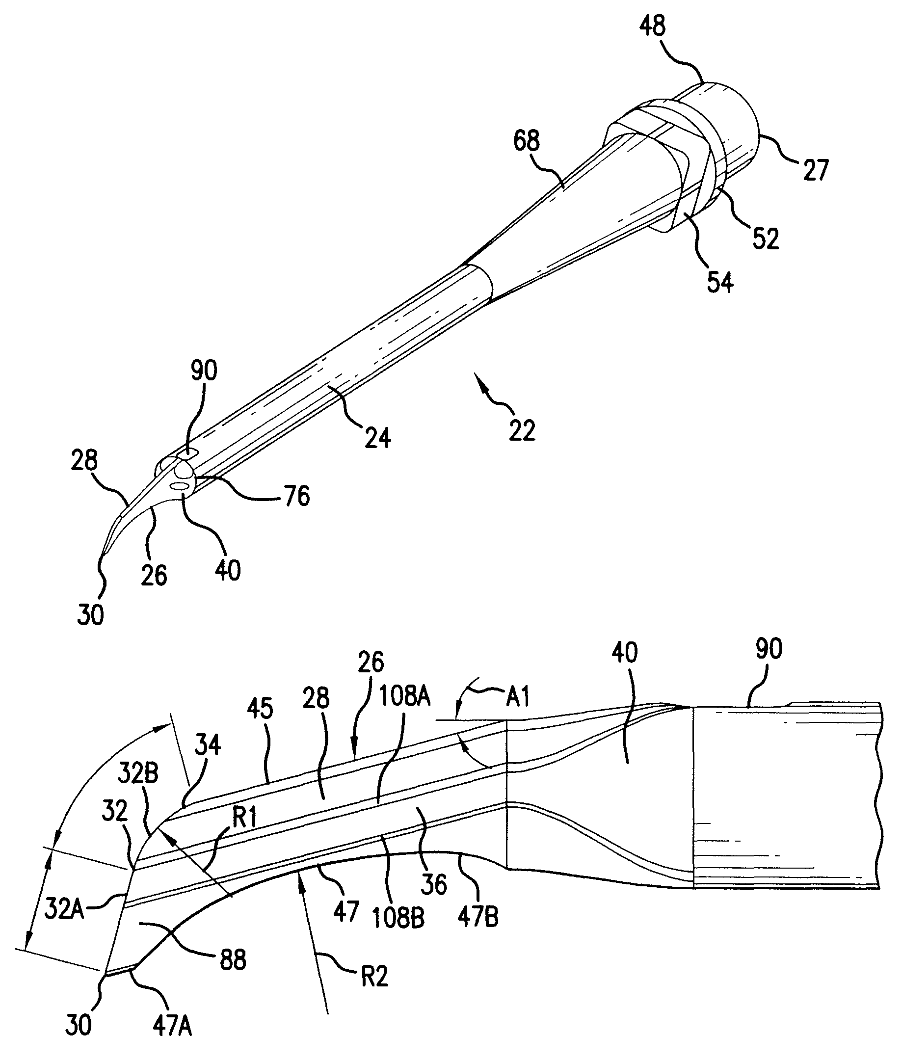
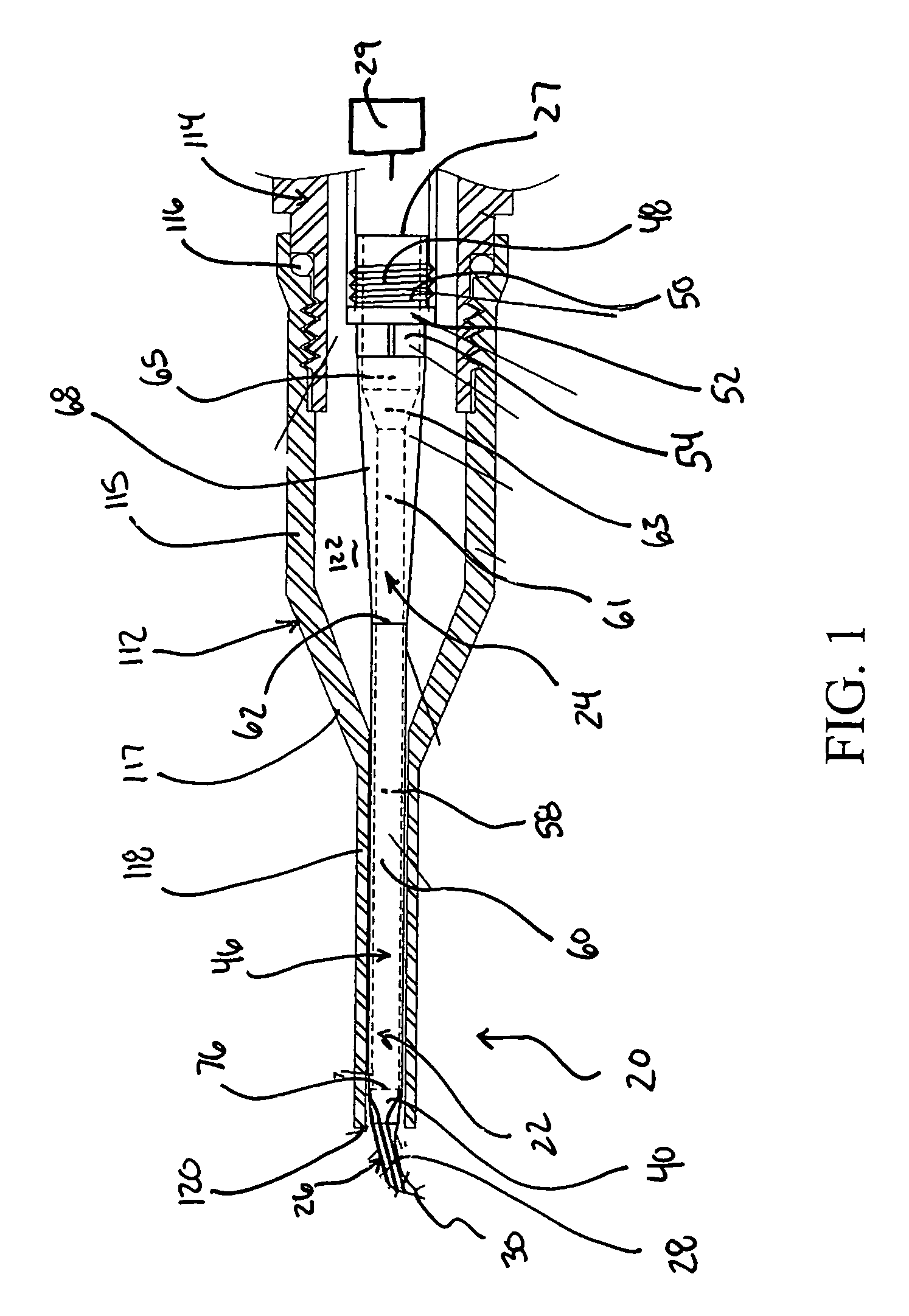
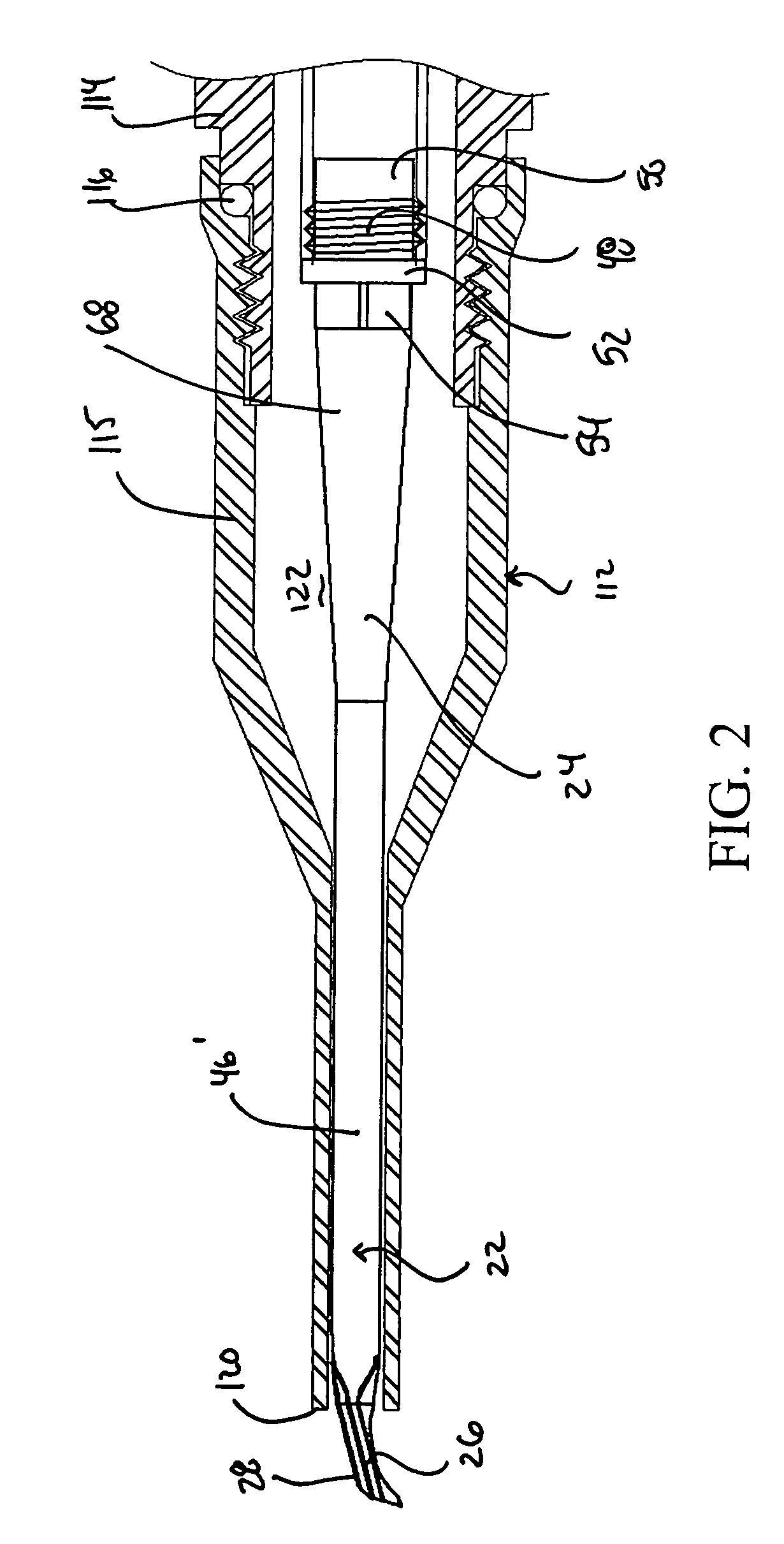
[0074]In a preferred embodiment, energy transmission section 24 includes shaft 46 (FIG. 1 featuring a hollow or internalized aspirating passageway embodiment, and FIG. 2 featuring a solid shaft or non-aspirating embodiment 46′) as well as connector 48 (or means for attachment) to a movement generator (schematically shown at 29 which is preferably in the form of an ultrasonic vibrating device such as one utilizing a piezoelectric transducer, although alternate movement generating devices are also featured under the present invention including, for example, alone or in conjunction with one or the other, fluid, acoustic, motor (e.g., offset cam), reciprocating piston or other movement generating means.

[0075]In a preferred embodiment shaft 46 includes a cylindrical shaft segment 60 that is preferably approximately 14 mm in length and 1 mm in diameter, although a variety of alternate cross-sectioned shaped, preferably straight shaft segments and lengths are featured under the subject mat...

second embodiment

[0098]With reference to FIGS. 12, 15 and 19 there is described cutting device 722 under this second embodiment set which is well suited for cutting in quick and efficient fashion through softer cataracts (e.g., non-black and non-brunescent cataracts).

[0099]As seen from FIG. 15, for example, cutting device 722 features a tip 724 with modified blade 726 extending off of a transition section 40 similar to that described above for the FIG. 1 embodiment. The height H2 (FIG. 19) of blade 726 is preferably the same as H1 (FIG. 8A) with a range of 0.7 to 2.0 mm being well suited for the preferred usages of the present invention. With reference to FIGS. 15 and 19 there can be seen forward, flat edge 732 having an upper end point 728 and a lower (material contact) point 730. The opposite walls 733 and 734 of blade 726 preferably converge from thicker upper edge 736 to lower edge 738 in similar fashion of the convergence discussion above (both vertically and axially), but with a preferred uppe...


PUM


Abstract

An ophthalmologic cutting device having a base support section for attachment with a movement generating device and a tip with a blade section. The blade section preferably has upper and lower edges, and a forward aspiration free edge extending between them, with the upper edge having a shorter longitudinal length compared with the lower edge and where the forward edge slopes down from a distal end of the upper edge to a distal end of the lower edge, and the lower edge presenting a material contact surface that is thinner in thickness than the upper edge. A slope back in the proximal direction of the forward edge of, for example, 10 to 45 degrees with a straight and / or curving forward edge or a combination of a straight and forward edge sections is preferred. Embodiments of the blade include a blade converging in thickness from top to bottom and one having a curved upper forward edge region and one with a lower edge that has a distal straight section and a recessed section positioned proximal of said distal straight section. The forward edge is also preferably defined by longitudinally diverging, opposing side walls.

Description

CROSS REFERENCE TO RELATED APPLICATIONS[0001]The present application claims priority under 35 USC §119(e) to U.S. Provisional Patent Application 60 / 715,800, filed Sep. 9, 2005, which is incorporated herein by reference in its entirety.FIELD OF THE INVENTION[0002]The invention relates to an ultrasonic cutting device that, in a preferred embodiment, is used as a cutting device in ophthalmologic surgery. The invention also pertains to surgical instruments and techniques comprising the ultrasonic cutting device. In a preferred embodiment the surgical instrument is a phacoemulsification ophthalmologic surgical instrument and the cutting device is a phaco tip for use in that instrument which provides a cutting device that is highly efficient in cutting cataracts into more manageable fragments.BACKGROUND OF THE INVENTION[0003]Phacoemulsification is currently the preferred technique used by eye surgeons for cataract extraction involving the removal of the cloudy eye crystalline lens. Throug...

Claims


Application Information

Patent Timeline
Patent Type & Authority Patents(United States)
IPC IPC(8): A61F9/007
CPCA61F9/00745A61B2017/320076A61B2017/320078A61B17/32A61B17/3209A61F9/007
Inventor ESCAF, LUIS J.
Owner ALCON INC
Why Eureka
Try Eureka