High-resolution single photon planar and spect imaging of brain and neck employing a system of two co-registered opposed gamma imaging heads

a gamma imaging head and co-registered technology, applied in the field of spect imaging, can solve the problems of large absorption of characteristic gamma radiation from tc-99m, availability and cost,

Active Publication Date: 2011-12-06
JEFFERSON SCI ASSOCS LLC
View PDF5 Cites 8 Cited by
  • Summary
  • Abstract
  • Description
  • Claims
  • Application Information

AI Technical Summary

Benefits of technology

[0011](1) The field of view coverage is significantly improved (about 20 cm×15 cm) over conventional SPECT brain imagers by providing two identical compact light-weight gamma imaging detector heads mounted on a rotating gantry and precisely mechanically co-registered to each other at 180 degrees (placed opposite to each other). The detector heads are placed on both sides of the patient and mechanically affixed relative to each other, with the patient's brain or neck fully encompassed by the resulting active field of view coverage.
[0012](2) The parallel-hole collimators mounted on the two gamma cameras are precisely mechanically aligned, with accuracy of alignment validated with point and linear radioactive sources.
[0016](6) The patient can be lying down on a bed, sitting straight in a chair, or optimally sitting in a chair in a reclined position in a chair to limit movements of the patient's head while providing patient comfort.
[0017](7) The patient's head will be attached to the imaging bed or chair to minimize head movements during scan but not to cause large discomfort.
[0018](8) The imaging bed, chair and / or imager gantry will allow for positioning of the central imaging plane at the proper height at the neck / head region to provide optimized imaging of the region of interest.
[0019](9) Following new imaging standards in high resolution imaging, position of the patient's head is constantly monitored by an optical monitoring system to correct the SPECT data for head movements during the scan.
[0020](10) The two compact detector heads can employ an array of compact position sensitive PMTs, but also the novel silicon photomultipliers (SiPMTs) making the system even more compact and potentially even more robust.
[0021](11) Implementation of SiPMTs and providing MRI-compatible shielding, collimators and other materials, which are immune to strong magnetic fields, permit insertion of the imager into an MRI magnet to achieve simultaneous dual-modality imaging, including fMRI-SPECT which combines two functional imaging modalities to study brain function.
[0022]These and other objects and advantages of the present invention will be better understood by reading the following description along with reference to the drawings.

Problems solved by technology

However, at the current time PET's availability and cost are obstacles to fast widespread introduction while single photon labeled biomarkers can be made available in most practical situations due to their longer half-lives, no need for nearby production centers, and lower cost.
The major problem associated with the single photon imaging procedure is that the characteristic gamma radiation from Tc-99m undergoes substantial absorption when traversing tissues such as brain.

Method used


Image

  • High-resolution single photon planar and spect imaging of brain and neck employing a system of two co-registered opposed gamma imaging heads
  • High-resolution single photon planar and spect imaging of brain and neck employing a system of two co-registered opposed gamma imaging heads
  • High-resolution single photon planar and spect imaging of brain and neck employing a system of two co-registered opposed gamma imaging heads

Examples


Embodiment Construction

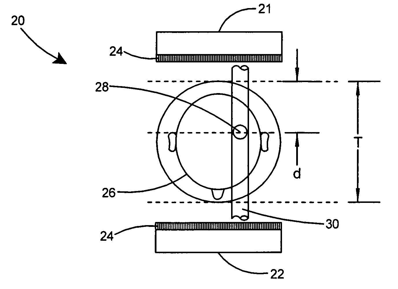
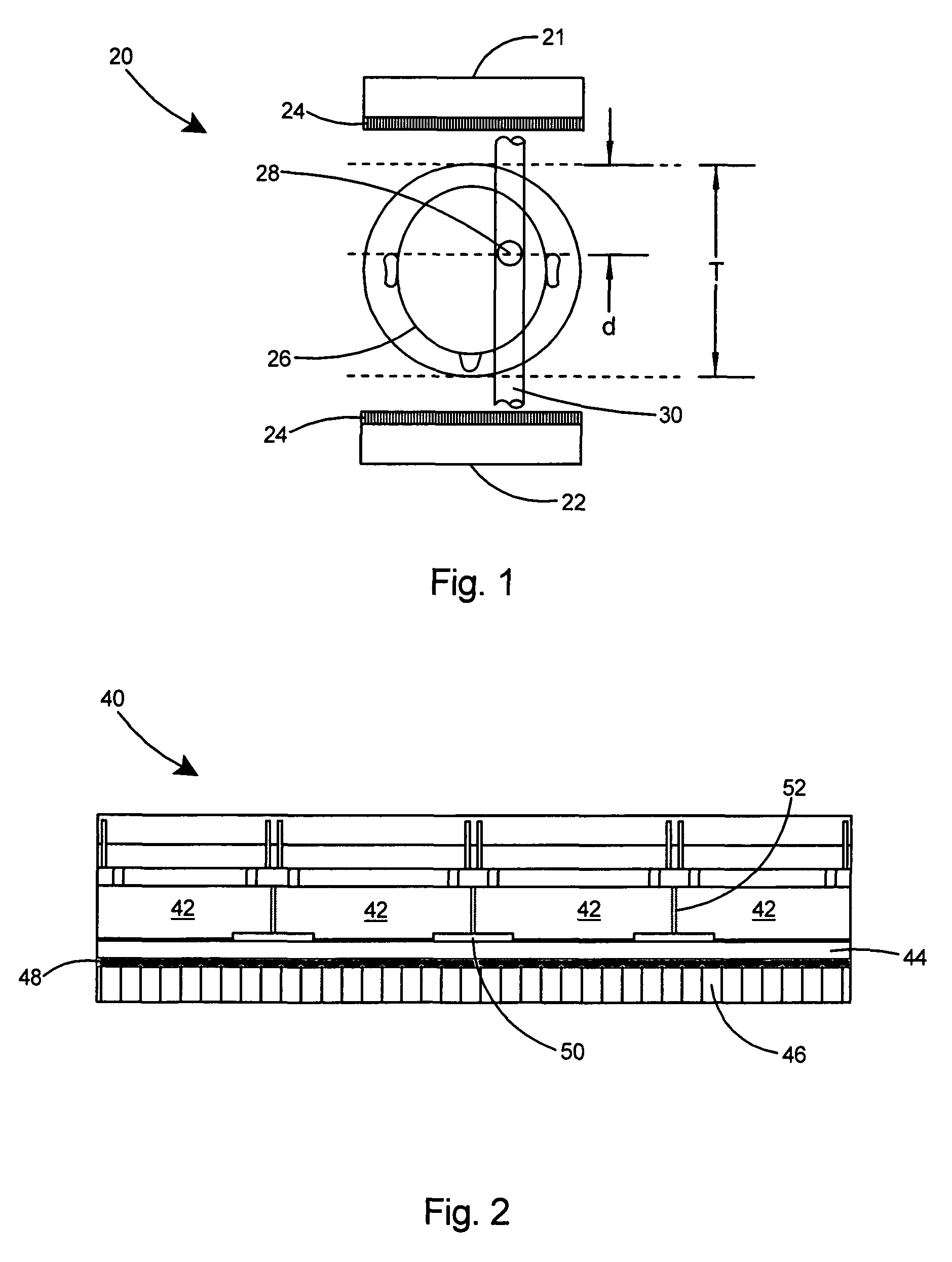
[0050]During conventional SPECT imaging, a series of images or projections are acquired from different angular detector head positions. Imaging heads are mounted on a rotating gantry. Each sector of the patient's head or neck has better visibility from the directions of closest approach to that sector, with the exception of the inner sectors of the head or neck that are equally distant from all directions. The imaging signal in the gamma camera from the inner sectors is much more attenuated and as a result image reconstruction for those sectors is poor.

[0051]A substantial improvement can be obtained in SPECT by employing the concept of combining projection images for each angular view from two opposed and co-registered gamma imaging heads, before implementing 3D tomographic reconstruction algorithm of the object. In addition, computer modeling of the absorption and spatial resolution effects, as well as subsequent correction of the collected data, would improve the quality of recons...


PUM


Abstract

A compact, mobile, dedicated SPECT brain imager that can be easily moved to the patient to provide in-situ imaging, especially when the patient cannot be moved to the Nuclear Medicine imaging center. As a result of the widespread availability of single photon labeled biomarkers, the SPECT brain imager can be used in many locations, including remote locations away from medical centers. The SPECT imager improves the detection of gamma emission from the patient's head and neck area with a large field of view. Two identical lightweight gamma imaging detector heads are mounted to a rotating gantry and precisely mechanically co-registered to each other at 180 degrees. A unique imaging algorithm combines the co-registered images from the detector heads and provides several SPECT tomographic reconstructions of the imaged object thereby improving the diagnostic quality especially in the case of imaging requiring higher spatial resolution and sensitivity at the same time.

Description

[0001]The United States of America may have certain rights to this invention under Management and Operating contract No. DE-AC05-06OR23177 from the Department of Energy.FIELD OF THE INVENTION[0002]This invention relates to SPECT imaging and more particularly to a compact and mobile high resolution SPECT imaging system for imaging the head, brain, and neck areas of a patient.BACKGROUND OF THE INVENTION[0003]Brain disease is becoming more prevalent in the aging population because of increased life span and Alzheimer's, Parkinson's, other dementias, and brain cancer will increase as US and European populations continue to age. Approximately 4,000,000 people in the U.S. have Alzheimer's disease. The US alone is expected to have 15 million Alzheimer's cases by 2050. Also an estimated 1.5 million people in the US have Parkinson's. Neurological brain disease affects 1% of Americans over 60 and each year there are 100,000 new cases. Diagnostic analysis of brain function represents an area o...

Claims


Application Information

Patent Timeline
Patent Type & Authority Patents(United States)
IPC IPC(8): G01T1/166
CPCA61B6/037A61B6/4266A61B6/501G01T1/1641A61B6/4417A61B6/583
Inventor MAJEWSKI, STANISLAWPROFFITT, JAMES
Owner JEFFERSON SCI ASSOCS LLC
Why Eureka
Try Eureka