Compositions consisting essentially of TGF-β, BMP-2 FGF-4, leukemia inhibitory factor, IGF-1, IL-6 and H-α-thrombin

a technology of tgf- and bmp-2 fgf-4, which is applied in the direction of drug compositions, peptides, cardiovascular disorders, etc., can solve the problems of cardiac remodeling and consequent impairment of myocardial function, impaired myocardial function, and limited mitotic capacity of cardiomyocytes to support adequate myocardial regeneration, etc., to achieve the effect of improving contractile performan

Active Publication Date: 2012-05-08
MAYO FOUND FOR MEDICAL EDUCATION & RES
View PDF10 Cites 9 Cited by
  • Summary
  • Abstract
  • Description
  • Claims
  • Application Information

AI Technical Summary

Benefits of technology

[0007]This document provides methods and materials related to treating cardiovascular tissue (e.g., heart tissue or vascular tissue). For example, this document provides stem cells, compositions containing stem cells, methods for obtaining stem cells, compositions for generating stem cells expressing particular markers, and methods for repairing cardiovascular tissue. The stem cells and compositions containing stem cells provided herein can be used to repair cardiovascular tissue. Such stem cells can allow clinicians to treat, for example, myocardial injury since the stem cells can have the ability to differentiate into cardiomyocytes. In some cases, such stem cells and compositions containing stem cells can be used to repair cardiovascular tissue without producing tumors within the tissue. The compositions provided herein for generating stem cells can allow medical professionals to produce large amounts of stem cells that can express particular markers and can be used to repair cardiovascular tissue.

Problems solved by technology

Myocardial injury leads to cardiomyocyte loss, ventricular remodeling and consequent impairment of myocardial function.
Unfortunately, the mitotic capacity of cardiomyocytes is too limited to support adequate myocardial regeneration.
Injecting 3300 cells per 1 mg of heart tissue can result in a 15 percent tumor risk, while injecting 10,000 cells per 1 mg of heart tissue can result in a 72 percent tumor risk.

Method used


Image

  • Compositions consisting essentially of TGF-β, BMP-2 FGF-4, leukemia inhibitory factor, IGF-1, IL-6 and H-α-thrombin
  • Compositions consisting essentially of TGF-β, BMP-2 FGF-4, leukemia inhibitory factor, IGF-1, IL-6 and H-α-thrombin
  • Compositions consisting essentially of TGF-β, BMP-2 FGF-4, leukemia inhibitory factor, IGF-1, IL-6 and H-α-thrombin

Examples


example 1

Materials and Methods for Embryonic Stem Cell Therapy in Myocardial Infarction

[0053]Embryonic stem cells. The CGR8 murine embryonic stem cell line was propagated in BHK21 or Glasgow MEM medium supplemented with pyruvate, non-essential amino acids, mercaptoethanol, 7.5% fetal calf serum, and the leukemia inhibitory factor (Meyer et al., 2000, FEBS Lett., 478:151-158; Perez-Terzic et al., 2003, Circ. Res., 92:444-452). A CGR8 cell clone was engineered to express the yellow fluorescent protein (YFP) or the enhanced cyan fluorescent protein (ECFP) tinder the control of the cardiac-specific α-actin promoter subcloned upstream of ECFP using XhoI and HindIII restriction sites of the promoterless pECFP vector (Clontech). This α-actin promoter construct was linearized using XhoI, and electroporated into CGR8 stem cells as described (Behfar et al., 2002, FASEB J., 16:1558-1566; Meyer et al., 2000, FEBS Lett., 478:151-158). To image stem cells by field-emission scanning electron microscopy, ce...

example 2

Embryonic Stem Cell Therapy in Myocardial Infarction

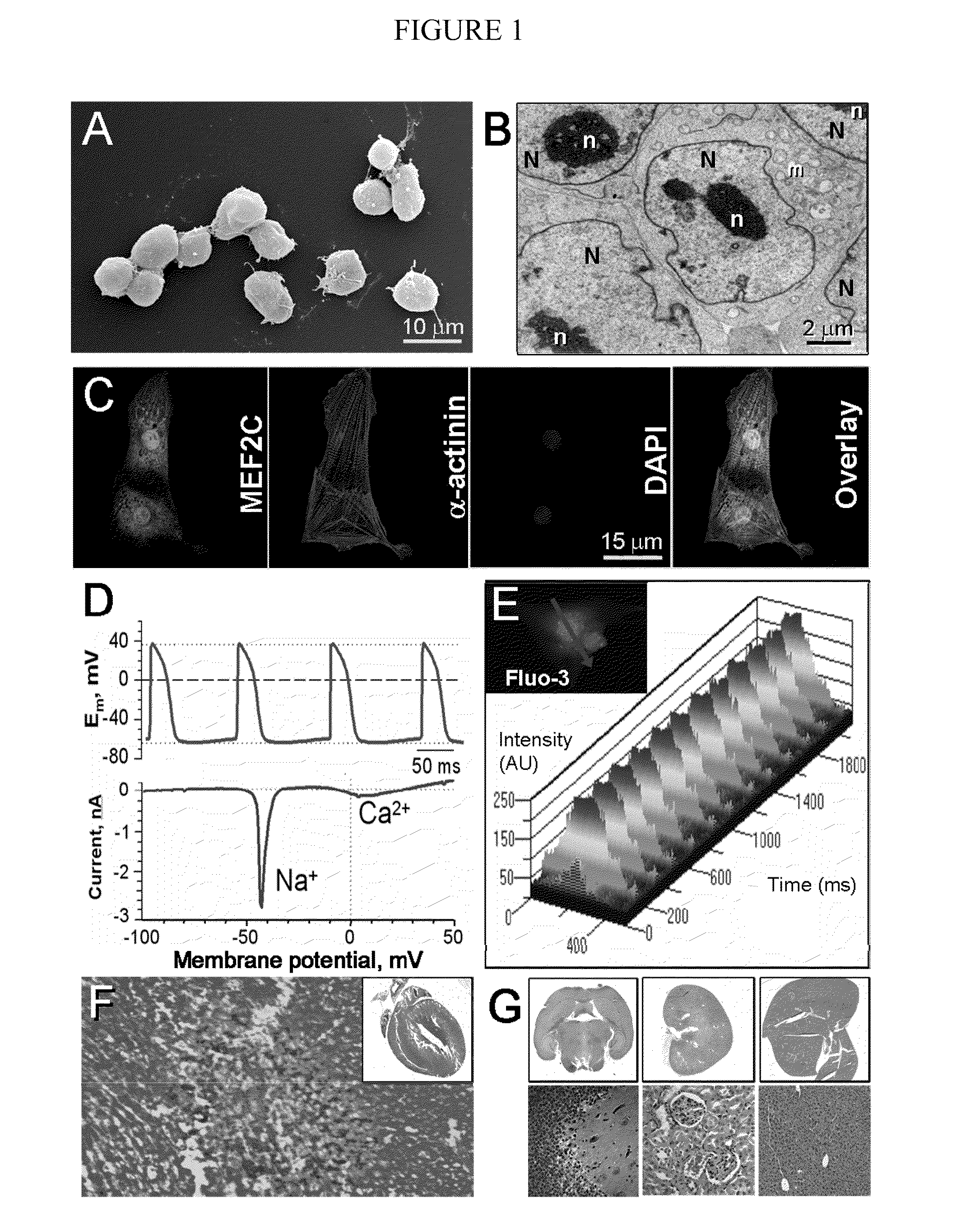
[0060]Cardiogenic potential of embryonic stem cells and delivery into infarcted heart. The CGR8 embryonic stem cell colony, used herein (FIG. 1A), demonstrated typical features of undifferentiated cells including a high nucleus to cytosol ratio, prominent nucleoli and mitochondria with few cristae (FIG. 1B). The cardiogenic capacity of this embryonic stem cell line was probed by in vitro differentiation, with cells readily derived that express the cardiac transcription factor MEF2C (Lin et al., 1997, Science, 276:1404-1407), the cardiac contractile protein α-actinin and sarcomeric striations (FIG. 1C). Consistent with proper differentiation towards cardiac lineage, stem cell-derived cardiomyocytes demonstrated action potential activity associated with prominent inward Na+ and Ca2+ currents (FIG. 1D), critical for excitation-contraction coupling manifested as rhythmic intracellular Ca2+ transients (FIG. 1E). Injection of CGR8 cells ...

example 3

Materials and Methods for Allogenic Stem Cells Dosed to Secure Cardiogenesis

[0066]Embryonic stem cells and derived cardiomyocytes. Field-emission scanning electron microscopy was used to visualize murine embryonic stem cells in culture (Perez-Terzic et al., 2003, Circ. Res., 92:444-452; Hodgson et al., 2004, Am. J. Physiol., 287:H471-H479). Differentiation in vitro was achieved using the hanging-drop method to generate embryoid bodies from which, following enzymatic dissociation, a highly enriched population of cardiomyocytes was isolated with a density gradient protocol (Behfar et al., 2002, FASEB J., 16:1558-1566; Perez-Terzic et al., 2003, Circ. Res., 92:444-452). Expression of cardiac markers was probed by laser confocal microscopy, with action potential activity captured by the patch-clamp method in the current clamp mode (Hodgson et al., 2004, Am. J. Physiol., 287:H471-H479).

[0067]Stem cell transplantation into host heart. To track transplanted cells in vivo, embryonic stem ce...


PUM

PropertyMeasurementUnit
pHaaaaaaaaaa
voltagesaaaaaaaaaa
pHaaaaaaaaaa

Abstract

This document provides methods and materials for treating cardiovascular tissue. For example, stem cells, compositions containing stem cells, methods for obtaining stem cells, compositions for generating stem cells expressing particular markers (e.g., compositions comprising TGF-β1, BMP-2, FGF-4, and leukemia inhibitory factor), and methods for repairing cardiovascular tissue are provided.

Description

CROSS-REFERENCE TO RELATED APPLICATIONS[0001]This application is a National Stage application under 35 U.S.C. §371 and claims benefit under 35 U.S.C. §119(a) of International Application No. PCT / US2005 / 026800 having an International Filing Date of Jul. 29, 2005, which claims the benefit of priority under 35 U.S.C. §119(e) of U.S. Application No. 60 / 680,775, filed May 12, 2005 and U.S. Application No. 60 / 592,871, filed Jul. 30, 2004. The disclosures of the prior applications are incorporated by reference in their entirety.BACKGROUND[0002]1. Technical Field[0003]This document relates to methods and materials involved in treating cardiovascular tissue with, for example, stem cells.[0004]2. Background Information[0005]Myocardial injury leads to cardiomyocyte loss, ventricular remodeling and consequent impairment of myocardial function. Unfortunately, the mitotic capacity of cardiomyocytes is too limited to support adequate myocardial regeneration. In addition, current therapeutic modali...

Claims


Application Information

Patent Timeline
Patent Type & Authority Patents(United States)
IPC IPC(8): A61K38/18C07K14/51C07K14/50C07K14/54C07K14/475C07K14/745C07K14/65A61K38/19C07K14/495A61K35/545
CPCA61K38/1825A61K38/1841A61K38/1875A61K38/204A61K38/2093A61K38/30A61K35/545A61K38/4833A61K2300/00A61P9/00
Inventor TERZIC, ANDREBEHFAR, ATTA
Owner MAYO FOUND FOR MEDICAL EDUCATION & RES
Why Eureka
Try Eureka