Method for enriching rare cell subpopulations from blood

a rare cell and blood technology, applied in the field of rare cell enrichment, can solve the problems of difficult to detect rare ctcs, and insufficient cell yield of current recovery methods, so as to increase the cellular recovery of non-erythrocytes, the effect of high sensitive and reproducibl

Active Publication Date: 2012-09-11
MEDICAL DISCOVERY PARTNERS
View PDF30 Cites 10 Cited by
  • Summary
  • Abstract
  • Description
  • Claims
  • Application Information

AI Technical Summary

Benefits of technology

[0015]In general, a method for enriching a cell population in a cell suspension includes removing cells that are not members of the cell population from the cell suspension. For example, a desired cell subpopulation can be isolated from blood, for analysis and possible use in clinical diagnosis. In certain circumstances, the method can be adapted to enrich cell subsets from a cell suspension, such as, for example, bodily fluids including blood, even when the cell subsets include a small fraction of the total cells in the suspension. The cell population can include rare cells. The cell suspension can be, for example, a fluid containing a mixture of rare cells and non-rare cells. The rare cells can be blood cells or non-blood cells. The rare non-blood cells can be, for example, epithelial cancer cells or cell types other than cancer cells. The cell population that is enriched can be a desired cell subpo

Problems solved by technology

Detecting rare CTCs is technically challenging.
Identifying and quantifying circulating carcinoma cells (malignant cells of epithelial origin) has been a technically challenging undertaking.
The currently available methods for recovering so few cells have insufficient cell yields (recoveries), often precluding rare cell isolation and subsequent analysis.
An important limitation of the ferrofluid technology is that it appears to not be effective as a cancer screening test.
According to the inventors of the Immunicon/Veridex technology, the limitation in sensitivity can be related to their positive selection method for CTC isolation.
This limits the utility of this widely-used marker for separating tumor cells from blood.
Another limiting factor is that many cells often begin to undergo apoptosis after detaching from the primary.
There are no FDA-cleared tests for CTCs using either method.
Cultured tumor cells generally are mostly buoyant on a

Method used


Image

  • Method for enriching rare cell subpopulations from blood
  • Method for enriching rare cell subpopulations from blood
  • Method for enriching rare cell subpopulations from blood

Examples


examples

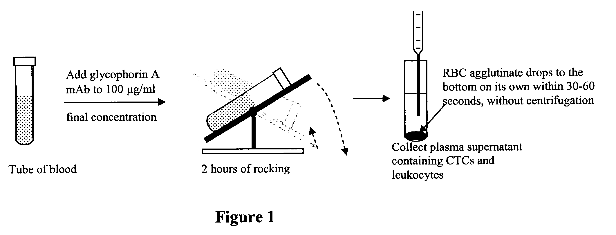
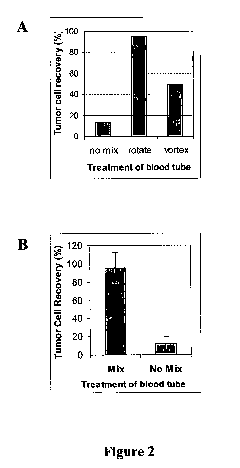
[0062]FIG. 2 illustrates the difference that mixing can make in tumor cell recoveries using blood specimens spiked with ZR-75-1 (breast carcinoma) tumor cells. Two separate experiments are shown (A and B). In the former (A), the tubes were either not mixed at all (“no mix”), mixed constantly on a rotator (“rotate”), or intermittently mixed on a vortex mixer (“vortex”). For the second experiment (B), the intermittent vortex mixing was not re-tested. The mixed group in experiment B used constant mixing on a tumbling shaker, whose motion is illustrated in the middle of FIG. 1. The vertical bar at left (experiment B) depicts tumor cell recovery with mixing (after adding the anti-glycophorin antibody). The right-hand bar demonstrates the tumor cell recovery for tubes left standing on the bench top during erythrocyte agglutination.

[0063]In these experiments, pathologic discard blood samples were procured from the Boston University Medical Center clinical laboratory, with approval and unde...


PUM


Abstract

An antigen-dependent negative selection blood cell separation method is described. Rare circulating epithelial cells can be separated from blood by depleting erythrocytes from a blood sample. Erythrocytes are depleted by agglutination. The new method comprises the use of an agglutinating agent, such as an anti-glycophorin A or glycophorin B antibody, as glycophorin A or B are present on erythrocytes and not on the desired epithelial cells. With regular mixing, desired rare circulating epithelial cells do not become entrapped in the red cell agglutinate.

Description

CLAIM OF PRIORITY[0001]This application claims priority under 35 U.S.C. §371 to International Application No. PCT / US2006 / 012595, filed on Apr. 4, 2006, which claims priority to U.S. Patent Application Ser. No. 60 / 669,388, filed Apr. 8, 2005, each of which is incorporated by reference in its entirety.TECHNICAL FIELD[0002]The present invention relates to a method of enriching rare cells from a fluid containing a mixture of rare cells and non-rare cells.BACKGROUND[0003]There has been growing interest in developing methods to detect and quantify circulating tumor cells from blood. Most types of cancer, such as tumors of the breast, prostate, colon, pancreas, esophagus, stomach, and liver are of epithelial origin. Blood cells, on the other hand, are of mesenchymal origin. This difference facilitates the detection of cancer cells, since epithelial-specific markers can be used to identify putative cancer cells in blood. Blood cells will not bear these epithelial-specific markers since they...

Claims


Application Information

Patent Timeline
IPC IPC(8): C12N5/00C12N5/02
CPCC12N5/0087G01N33/5091G01N33/56966G01N33/574G01N33/80
Inventor OLKEN, SARAH K.BOGEN, STEVEN A.SOMPURAM, SESHI R.
Owner MEDICAL DISCOVERY PARTNERS
Why Eureka
Try Eureka