Overlay image contrast enhancement
a contrast enhancement and overlay technology, applied in image enhancement, instruments, applications, etc., can solve the problems of image dissimilarity and difficult comparison
- Summary
- Abstract
- Description
- Claims
- Application Information
AI Technical Summary
Benefits of technology
Problems solved by technology
Method used
Image
Examples
example 1
[0149]Embodiments of the invention comprise a method for the imaging of contrast agents for high-resolution, high-frequency ultrasound imaging of animal subjects including rodent models.
[0150]As described above, contrast agents can flow in the blood stream of the animal and can be deposited at any site that the blood flows. Contrast agents can also be prepared in such a way that they can attach themselves to specific markers in the animal. This “targeted” contrast agent technique uses ligands and antibodies to latch onto specific targets. Contrast agents can also be used for image enhancement to allow a user to easily distinguish small vessels in the organ. In one aspect, perfusion imaging is a way of injecting contrast agent into the animal, allowing the agent to circulate and then the agent can be destroyed using a destruction event. The time taken for the re-perfusion is meaningful to the researcher in quantifying perfusion. This image processing method is not specific to the ima...
example 2
Materials and Methods
[0175]Inflammation in the mouse hindleg was induced by a one or three hour treatment with TNF-alpha injected subcutaneously into the hindpaw. Inflammation in the kidney was induced by ischemia-reperfusion injury. The left kidney was exposed and the renal artery was clamped for 32 minutes, followed by 2 hours of re-perfusion as described in Singbartl K, Green S A, Ley K. (2000) “Blocking P-selectin protects from ischemia / reperfusion induced acute renal failure”FASEB J. 14: 48-54. The wound was closed in layers and, covered with a saline-soaked gauze.
[0176]TargestarB microbubbles from Targeson (Charlottesville, Va.) were targeted to P-selectin by conjugating an anti-P-selectin monoclonal antibody to the surface of the MB per manufacturer's instructions. The MB were diluted to a concentration of 107 or 108 MB in 100 μL of phosphate-buffered saline, and injected as a bolus through a cannula placed in the left jugular vein.
[0177]Ultrasound imaging was performed using...
example 3
Image Enhancement by Bolus Injection of Vascularized Tissue
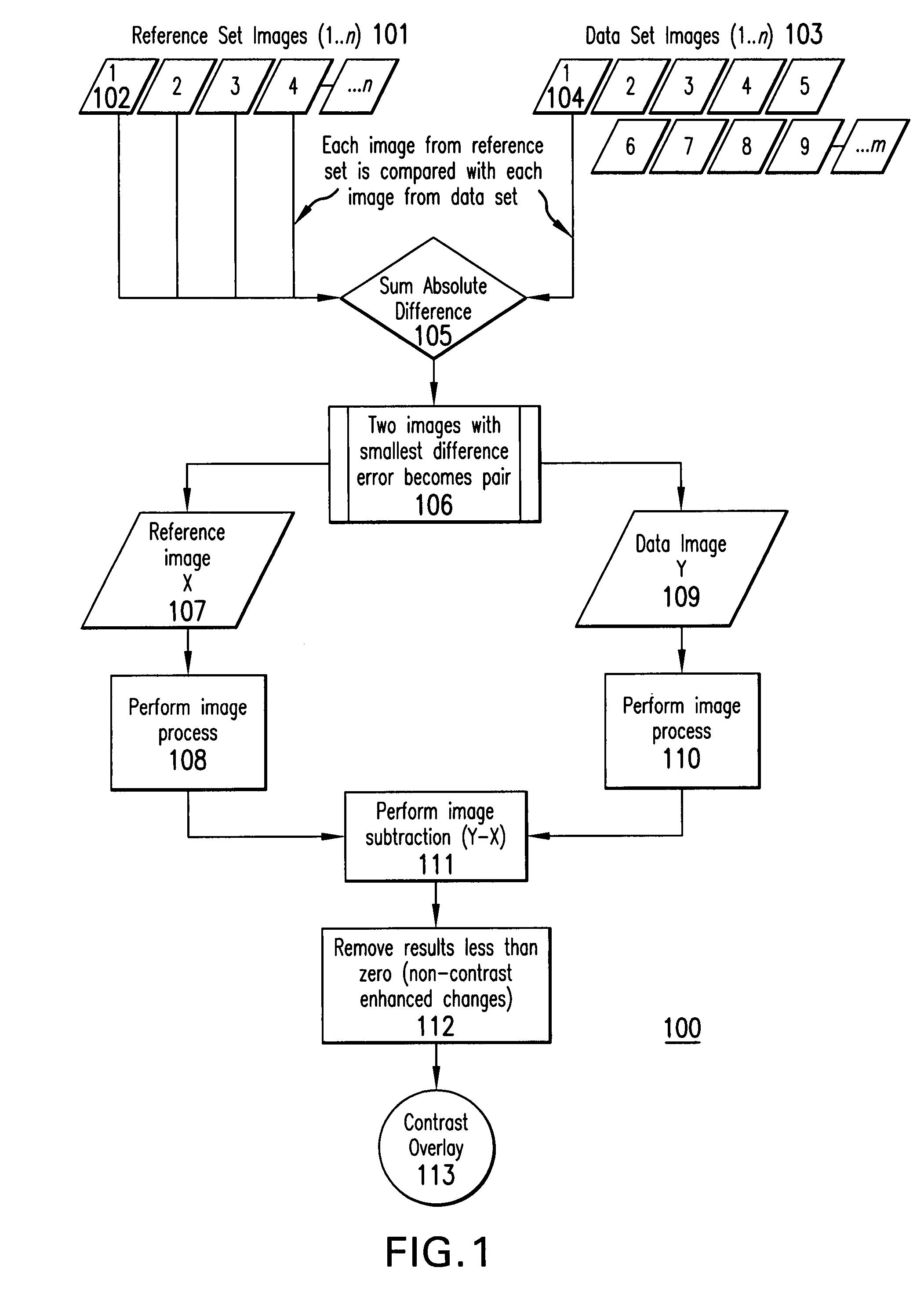
[0185]A Vevo 770® (VisualSonics Inc. Toronto, Calif.) high-resolution imaging system was used to acquire data set images and reference set images. The Vevo® system included software for comparing, matching and subtracting data set images and reference set images. For example, Vevo® Contrast Mode software was used.
[0186]The Default Cine Loop Size for Contrast Mode was set at 800, which defined the size of the Contrast Mode cine loop. A high frequency ultrasound imaging probe was connected to the to the Vevo® 770 imaging system. In this example, an RMV™-706 probe (VisualSonics Inc., Toronto, Calif.) was used.
[0187]The subject was positioned for scanning, and while scanning, the Field of View was adjusted to be 9×9 mm. Images were acquired at a frame rate between 10 and 20 Hz and the transmit power was set to 50%.
[0188]Contrast agent was prepared according to the instructions provided in VisualSonics, Inc. (Toronto, Calif.) Pre...
PUM
 Abstract
Description
Claims
Application Information
 - R&D
- Intellectual Property
- Life Sciences
- Materials
- Tech Scout
- Unparalleled Data Quality
- Higher Quality Content
- 60% Fewer Hallucinations
Browse by: Latest US Patents, China's latest patents, Technical Efficacy Thesaurus, Application Domain, Technology Topic, Popular Technical Reports.
© 2025 PatSnap. All rights reserved.Legal|Privacy policy|Modern Slavery Act Transparency Statement|Sitemap|About US| Contact US: help@patsnap.com


