X-ray pixel beam array systems and methods for electronically shaping radiation fields and modulation radiation field intensity patterns for radiotherapy

a radiation field and intensity modulation technology, applied in the field of x-ray radiation therapy, can solve the problems of age-adjusted cancer death rate in the united states that has not shown a corresponding improvement, tumors created from established cell lines implanted into non-native sites can respond differently, and the effect of radiation intensity and intensity modulation pattern

Active Publication Date: 2012-11-06
DUKE UNIV +1
View PDF15 Cites 64 Cited by
  • Summary
  • Abstract
  • Description
  • Claims
  • Application Information

AI Technical Summary

Problems solved by technology

Despite the significant advances in cancer diagnosis, treatment technologies, and research in recent years, the age-adjusted cancer death rate in the United States has not shown a corresponding improvement.
Further, quality of life issues have been receiving a growing amount of attention from cancer patients and healthcare providers.
However, the standard xenograft model of implanting tumor cells at various sites of immunocompromised mice has inherent limitations in that, while easily accessible for irradiation and measurement, tumors created from established cell lines implanted into non-native sites can respond differently than spontaneous tumors arising in their native environment.
In this case, however, monitoring tumor size by palpation is difficult or impossible for non-superficial sites.
Furthermore, delivery of radiotherapy for radiosensitization studies using orthotopic models is complicated by the radiation toxicity associated with many sites.
Targeted radiotherapy delivery, with confirmation of adequate treatment of the tumor while sparing adjacent normal tissues has been a primary limitation to such studies.
Often mouse irradiation intended for the tumor is also unavoidably given to the entire body or a large portion of it.
Consequentially severe radiation reactions are prone to develop in the mouse model that can greatly hinder the range of study possible.
Because of the lack of imaging devices in most animal model research laboratories, many orthotopic models with internal tumors that are not easily palpable cannot be used for irradiation study.
Conformal and fractionated irradiation, as used in human radiotherapy, is also not possible in current small animal irradiation technology.
Such technical limitations prevent researchers from fully using the animal models under conditions analogous to those used for human treatment, thereby weakening the clinical relevancy of the research.
There are considerable technical challenges in developing these proposed small animal irradiation and imaging devices to meet the spatial and temporal resolution demands of high quality small animal imaging and conformal irradiation.
One challenge is related to the mechanical complexity in the design, fabrication, and control of the miniaturized radiation field collimation system.
Assuming that the micro-RT system includes features similar to linear accelerators for human RT, that is, with the high accuracy and automatic collimator motion needed for intensity-modulated treatment, the miniature scale involved in micro-RT irradiation of small animals would be difficult to develop.
Another challenge is the temporal resolution required for high quality imaging and image-guided conformal irradiation of live small animals capable of rapid organ motion.
Conventional x-ray tubes deliver continuous radiation and thus can lead to motion-blurred images.
In addition, the radiation dose from imaging alone may be high.
Therefore, modern conventional clinical LINAC is very complicated mechanically and electronically, and the there are mechanical constraints on MLC motion (configuration and speed) so that some radiation field configurations and intensity patterns are not physically possible.
This dependence on mechanical means hinders the advancement of the RT technologies to meet the increasing demand to deliver RT treatments with higher spatial and temporal resolution for better treatment outcome.
The increasingly complex mechanical components and thus their electronic control systems can also significantly raise the cost of cancer treatment (device purchase cost and the ongoing maintenance cost include technical staff) and at the same time drop reliability of the RT machine.

Method used


Image

  • X-ray pixel beam array systems and methods for electronically shaping radiation fields and modulation radiation field intensity patterns for radiotherapy
  • X-ray pixel beam array systems and methods for electronically shaping radiation fields and modulation radiation field intensity patterns for radiotherapy
  • X-ray pixel beam array systems and methods for electronically shaping radiation fields and modulation radiation field intensity patterns for radiotherapy

Examples


Embodiment Construction

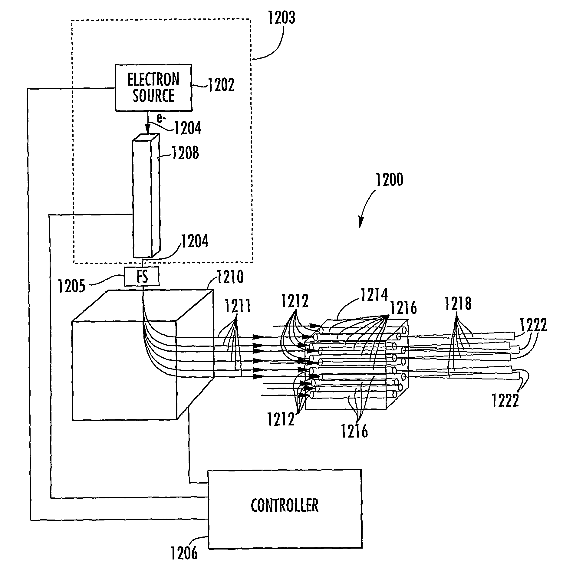
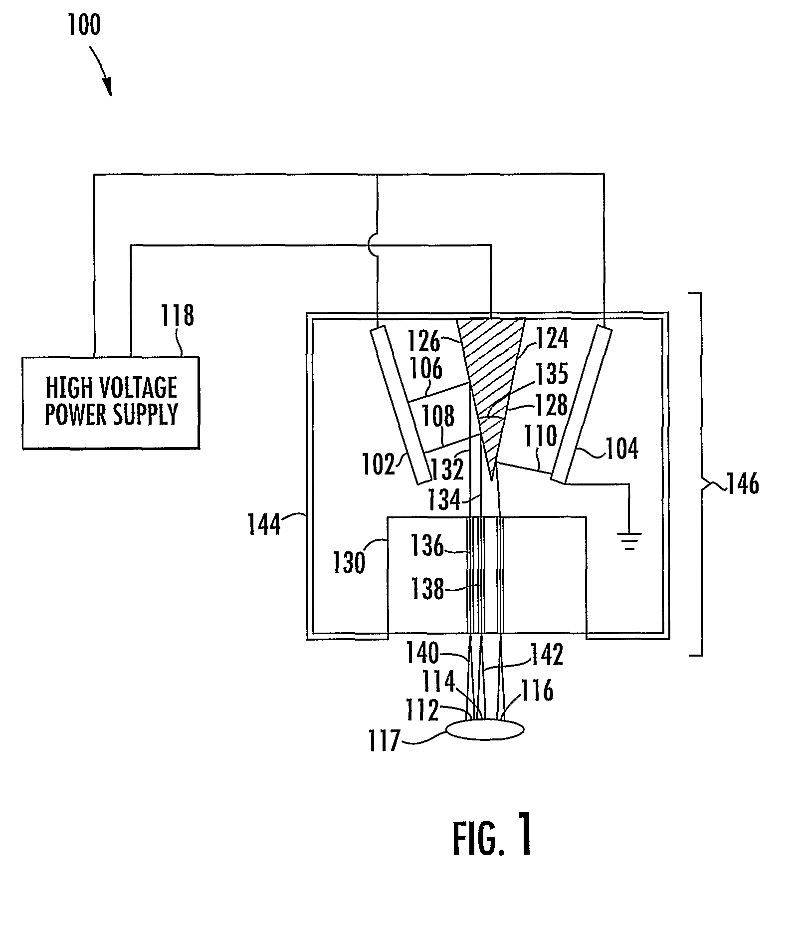
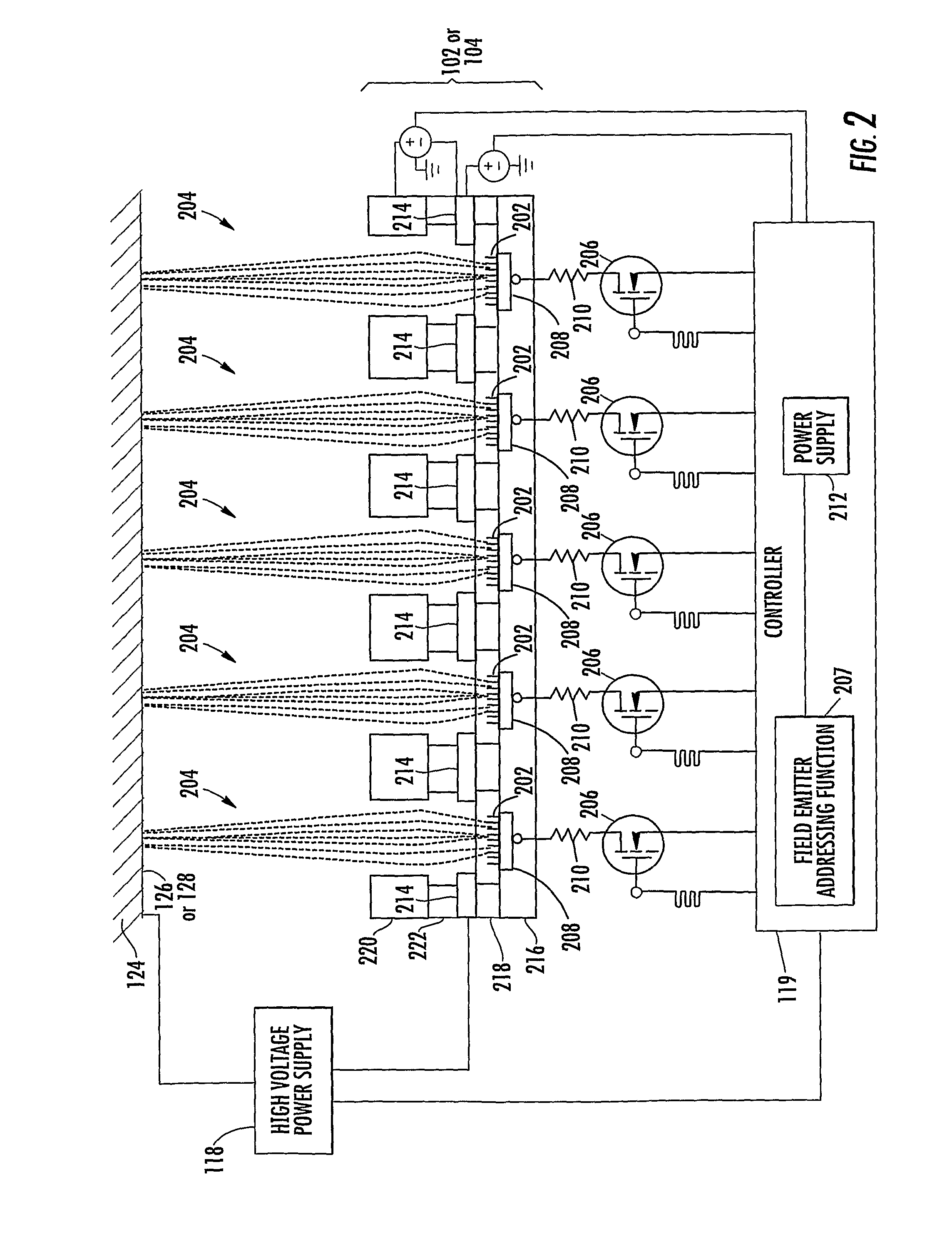
[0039]In accordance with the present disclosure, x-ray pixel array systems and related methods are provided. The systems and methods described herein may have particular application for use in electronically shaping radiation field and modulating radiation intensity pattern for subject irradiation. CT imaging and radiotherapy (RT) treatment are exemplary uses of the systems and methods described herein. In particular, the systems and methods described herein may be used for delivering radiation to animals in a pre-clinical setting and / or humans in a clinical setting.

[0040]For a pre-clinical application, an x-ray pixel array system according to the present disclosure may include a plurality of addressable electron field emitters operable to emit a plurality of electron pixel beams. Further, an x-ray pixel array system according to the present disclosure may include an anode positioned to accelerate electrons in the electron pixel beams and to convert a portion of energy associated wi...


PUM

PropertyMeasurementUnit
energyaaaaaaaaaa
energyaaaaaaaaaa
voltage potentialaaaaaaaaaa

Abstract

X-ray pixel beam array systems and methods for electronically shaping radiation fields and modulating radiation field intensity patterns for radiotherapy are disclosed. One exemplary pre-clinical system may include addressable electron field emitters (102, 104) that are operable to emit a plurality of electron pixel beams (106, 108, 110). Each electron pixel beam may correspond to an x-ray target (124) and x-ray pixel beam collimation aperture (136, 138) to convert a portion of energy associated with the electron pixel beam to a corresponding x-ray pixel beam (140, 142). Further, the x-ray pixel beam array collimator (130) forms a one-to-one correspondence between individual electron pixel beam and its corresponding x-ray pixel beam. One exemplary clinical system may include a high-energy electron source (1203), an n-stage scanning system (1210), x-ray pixel beam targets (1212), and an x-ray pixel beam array collimator (1214). A controller (1206) may sequentially direct electron beam pulses to predetermined x-ray pixel targets and produce an electronically controlled radiation field direction, pattern; and intensity pattern.

Description

CROSS-REFERENCE TO RELATED APPLICATION[0001]This non-provisional patent application claims the benefit of U.S. Provisional Application No. 60 / 685,791, filed May 31, 2005, the disclosure of which is incorporated by reference herein in its entirety.TECHNICAL FIELD[0002]The subject matter disclosed herein relates to using x-rays for radiotherapy treatment in clinical and pre-clinical use. More particularly, the subject matter disclosed herein relates to x-ray beam pixel array systems and methods for electronically shaping radiation fields and intensity modulation patterns for clinical and pre-clinical radiotherapy applications.BACKGROUND ART[0003]Many cancer patients receive radiotherapy (RT) during the course of cancer treatment. RT may be used alone or combined with other treatment modalities. Despite the significant advances in cancer diagnosis, treatment technologies, and research in recent years, the age-adjusted cancer death rate in the United States has not shown a corresponding...

Claims


Application Information

Patent Timeline
Patent Type & Authority Patents(United States)
IPC IPC(8): G01N23/04H05G1/70A61N5/10
CPCA61N5/103A61N2005/1061A61N5/1043A61N5/1077A61N2005/1095
Inventor CHANG, SHA X.WU, YING
Owner DUKE UNIV
Why Eureka
Try Eureka