Diagnostic test for renal injury

a technology for diagnosing renal injury and detecting the injury, applied in the direction of instruments, biochemistry apparatus and processes, material analysis, etc., can solve the problems of false positive diagnosis and other problems, and achieve the effects of satisfactory analytical specificity, sensitivity and precision, and greater diagnostic specificity

Active Publication Date: 2012-11-20
BIOPORTO DIAGNOSTICS AS
View PDF68 Cites 2 Cited by
  • Summary
  • Abstract
  • Description
  • Claims
  • Application Information

AI Technical Summary

Benefits of technology

[0044]An additional advantage of the present invention is that a cutoff value for the urine:plasma NGAL concentration ratios determined by one analytical technique can be applied to results obtained by a different analytical technique. This is because the concentration ratios obtained, unlike absolute concentration values, will be independent of the analytical techniques used and possible differences in their calibration, always provided that the analytical method shows a satisfactory linearity and is not influenced by differences in the composition of urine, plasma or serum that are not differences in the NGAL concentration.
[0045]The method of the present invention is particularly useful in patients admitted to intensive or critical care departments, or in any patient who is affected by a serious inflammatory, infective or neoplastic disease or disorder, which in itself or through its complications carries a high risk of causing renal injury leading to ARF or ARD. In this situation, the inflammatory, infective or neoplastic disorder may itself give rise to an elevated NGAL concentration before any renal injury has occurred. This elevation will usually be more marked in plasma than in urine, and the plasma level may in certain circumstances exceed the cutoff level above which the NGAL concentration in plasma is normally regarded as diagnostic of renal injury leading to ARD. The method of the present invention will not diagnose such a patient as having a renal injury leading to ARD until such time as the urinary concentration of NGAL rises to give a urine:plasma NGAL concentration ratio above the diagnostic cutoff value determined for this ratio, thus providing, in these patients, greater diagnostic specificity for renal injury leading to ARD than plasma or urinary levels of NGAL can separately provide.
[0046]The method of the present invention will also be useful in patients who, while not being critically ill, have suffered a well-defined insult such as a surgical operation that may lead to ischemic injury of the kidney or have been exposed to a nephrotoxic agent, such as a chemotherapeutic or antimicrobial agent or intravenously administered contrast medium for diagnostic imaging. In these cases the early identification of patients whose kidneys have been affected by the procedure and who are therefore at risk of ARF may allow for an earlier and more intensive intervention to prevent the development of ARF.
[0047]The method of the present invention will also be useful in veterinary medicine, the investigation of renal pathophysiology in experimental animals, and the testing of candidate therapeutic or diagnostic agents for nephrotoxicity in laboratory animals.
[0048]Measurement of NGAL in a sample of bodily fluid, such as a sample of urine, plasma or serum, can be performed by any method that provides satisfactory analytical specificity, sensitivity and precision. Preferred methods are binding assays using one or more binding molecules specific to NGAL and capable of binding to NGAL from the mammalian species from which the samples are obtained. Such binding molecules include, but are not limited to, polyclonal or monoclonal antibodies against NGAL or specific NGAL binding molecules generated by other means.
[0049]In a preferred method, monoclonal antibodies raised against recombinant NGAL from the mammalian species to be analyzed are used. One antibody is linked to a solid support to capture NGAL from a sample, such as a urine sample, a blood plasma or serum sample, while the other is labeled with at least one of a variety marker substances or chemical moieties, including but not limited to a chromophore, dye complex, fluorophore, electrochemical or chemoluminescence marker, or biotin or enzyme, that can be detected by any of many methods known to those skilled in the art. The solid support may e.g. be a polystyrene or polyvinyl chloride surface for enzyme-linked immunosorbent assay (ELISA), or latex (polystyrene) particles, or a filter frit composed of compressed polyethylene particles, or a porous nitrocellulose matrix, or indeed any suitable support used in immunochemical analyses. Particles coated with molecules that bind specifically to NGAL can also be used to determine NGAL concentrations in a sample by particle-enhanced turbidimetric or nephelometric methods, whether performed manually or by automated methods.

Problems solved by technology

When a cutoff level is used, these two types of borderline cases may give rise to false positive diagnoses of renal injury or false negative diagnoses of absence of renal injury, respectively.

Method used


Image

  • Diagnostic test for renal injury
  • Diagnostic test for renal injury
  • Diagnostic test for renal injury

Examples


example 1

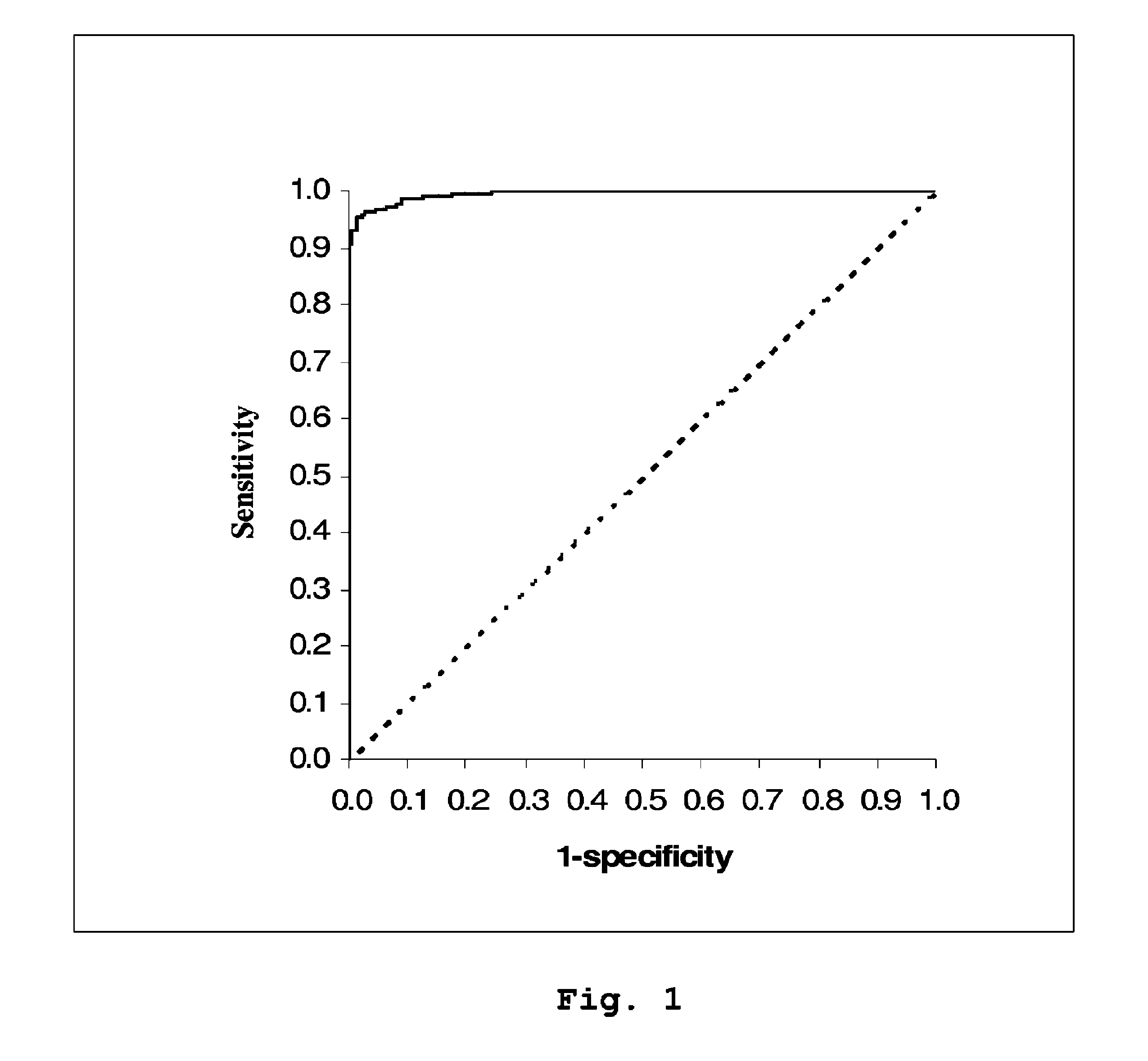
Diagnostic Power with Respect to ARF or ARD of the Urine:Plasma NGAL Concentration Ratio in 458 Paired Urine and Plasma Samples from 90 Adult Patients Admitted to Intensive Care

[0053]NGAL was determined (by means of an NGAL ELISA kit providing the sandwich ELISA described in Example 5) in 458 paired samples of urine and plasma collected each morning from a total of 90 patients admitted to a hospital intensive care unit. On the basis of discharge summaries and the results of routine blood tests, the patients were classified (blindly with respect to NGAL data) into those with one or more episodes of ARF or ARD during their admission (37 patients, giving 192 paired samples associated with episodes of ARF or ARD at the time of sampling or 24 hours later) and those without episodes of ARF or ARD (53 patients, 266 paired samples). The median urine:plasma NGAL concentration ratio in the sample pairs associated with an episode of ARF or ARD was 3.18 (range 0.33 to 40.02), while that in samp...

example 2

NGAL Dipstick Test

[0056]The analytical area of a dipstick comprised of a polystyrene surface is coated with a capture antibody against human NGAL. An aliquot of the centrifuged, diluted sample is added to a solution of enzyme-labeled detection antibody against NGAL in the first tube, into which the dipstick is immersed. Complexes of enzyme-labeled detection antibody with NGAL are bound to the dipstick, which is then washed with tap water and placed in a chromogenic substrate solution in a second tube. The color developed in the substrate solution within a given time is read either by eye and compared with a chart of color intensities which indicates the concentration of NGAL in the urine sample, or in a simple calorimeter that can, for example, be programmed to indicate the NGAL concentration directly.

example 3

NGAL Lateral Flow Device

[0057]A lateral flow device comprised of a strip of porous nitrocellulose or other material with channels capable promoting the migration of liquid by capillary forces is coated near its distal end with a capture antibody against NGAL applied as a transverse band. A further transverse band of antibody against antibodies of the species from which the detection antibody is derived is placed distally to the capture antibody band and serves as a control of strip function. The proximal end of the strip contains the detection antibody against NGAL adsorbed or linked to labeled polystyrene particles or particles of dye complex. When an aliquot of the centrifuged or filtered sample is applied to the proximal end of the strip, the labeled particles attached to detection antibody travel along the strip by capillary attraction. When reaching the band of capture antibody, only those particles which have bound NGAL in the sample will be retained, giving rise to a detectab...


PUM

PropertyMeasurementUnit
concentrationaaaaaaaaaa

Abstract

A method is provided of diagnosing and monitoring acute renal injury leading to acute renal failure in a human or mammalian subject by determining the ratio of the concentration of neutrophil gelatinase-associated lipocalin (NGAL) in urine to that in plasma or serum.

Description

[0001]This patent application is the National Stage of International Application No. PCT / DK2008 / 050069, filed Mar. 18, 2008, which claims the benefit of priority from U.S. Provisional Patent Application Ser. No. 60 / 919,277, filed Mar. 21, 2007, teachings of each of which are herein incorporated by reference in their entirety.FIELD OF THE INVENTION[0002]The present invention relates to the diagnosis and monitoring of a recently occurred or ongoing injurious process in a mammal that is affecting the kidneys and may lead to acute renal failure (ARF), hereinafter also synonymously referred to as acute renal dysfunction (ARD). As such it is relevant to internal medicine and in particular to critical or intensive care medicine, but also to surgery, oncology and diagnostic imaging, where procedures that may injure the kidneys are carried out. It is also relevant to veterinary medicine and surgery and to the study of renal pathophysiology and noxious influences on the kidney in laboratory a...

Claims


Application Information

Patent Timeline
Patent Type & Authority Patents(United States)
IPC IPC(8): C12Q1/37G01N33/493G01N33/50G01N33/53G01N33/573
CPCG01N33/6893G01N2800/347G01N2333/82
Inventor UTTENTHAL, LARS OTTOBANGERT, KRISTIAN
Owner BIOPORTO DIAGNOSTICS AS
Why Eureka
Try Eureka