Nested balloon catheter for localized drug delivery

a catheter and drug technology, applied in catheters, angiography, therapy, etc., can solve the problems of undesirable extravasation of diagnostic or therapeutic agents into tissue, many therapeutic and diagnostic many diagnostic and therapeutic agents in general may not be delivered using, etc., to facilitate the infusion of therapeutic and/or diagnostic agents

Active Publication Date: 2013-01-08
SANOVAS
View PDF75 Cites 27 Cited by
  • Summary
  • Abstract
  • Description
  • Claims
  • Application Information

AI Technical Summary

Benefits of technology

[0016]Accordingly, it is an object of the present invention to provide a nested balloon catheter system that can deliver therapeutic and / or diagnostic agents to bodily tissues, tumors, and other biological materials from within bodily cavities.
[0017]It is a further object of the present invention to provide a nested balloon catheter system that can target specific areas for the delivery of therapeutic and / or diagnostic agents to bodily tissues, tumors, and other biological materials.
[0018]It is yet another object of the present invention to provide a nested balloon catheter system that facilitates infusion of therapeutic and / or diagnostic agents into surrounding bodily tissues, tumors, and other biological materials.
[0019]It is still another object of the present invention to provide a nested balloon catheter system for delivering therapeutic and / or diagnostic agents to bodily tissues, tumors, and other biological materials that provides physiological feedback from which the intra-lumen diameter of the bodily cavity can be determined, and the pressure and flow supplied to the balloon can be adjusted accordingly.
[0029]In some embodiments, the catheter further includes a distal balloon positioned distally of the inner and outer balloons and a proximal balloon positioned proximally of the inner and outer balloons, wherein the fluid source inflates the proximal balloon and the distal balloon by supplying fluid thereto to create a chamber therebetween. In some of these embodiments, an outer surface of the proximal and distal balloons has a textured surface for preventing slippage of the outer surface on tissue.

Problems solved by technology

However, many therapeutic and diagnostic agents in general may not be delivered using these routes because they might be susceptible to enzymatic degradation or cannot be absorbed into the systemic circulation efficiently due to molecular size and charge issues, and thus, will not be therapeutically effective.
There are several known problems associated with the injection process.
One of such problems is undesirable extravasation of the diagnostic or therapeutic agents into tissue, which is particularly prevalent with intravenously injected agents.
Extravasation generally refers to leakage of fluids out of a container, and more specifically refers to leakage of intravenous drugs from a vein into surrounding tissues, resulting in an injury to the tissues.
Once the intravenous extravasation has occurred, damage can continue for months and involve nerves, tendons and joints.
If treatment is delayed, surgical debridement, skin grafting, and even amputation have been known to be the unfortunate consequences.
Occurrence of extravasation is possible with all intravenuous drugs, but it is a particularly significant problem with cytoxic drugs used for treatment of cancer (i.e. during chemotherapy).
Unfortunately, it cannot tell the difference between a cancer cell and some healthy cells.
With permanent implants (e.g. prostate), the radioactivity of the seeds typically decays with time.
Veins of people receiving chemotherapy are often fragile, mobile, and difficult to cannulate.
Patients who have had previous radiation therapy at the site of injection may develop severe local reactions from cytotoxic drugs.
Cytotoxic drugs also have the potential to cause cutaneous abnormalities in areas that have been damaged previously by radiation, even in areas that are distant from the injection site.
Patients who receive further chemotherapy in a different site may experience an exacerbation of tissue damage in the original site.
Furthermore, areas of previous surgery where the underlying tissue is likely to be fibrosed and toughened dramatically present an increased risk of extravasation.
Radical mastectomy, axillary surgery or lymph node dissection may impair circulation in a particular limb.
Some chemotherapy drugs often never reach the tumors they are intended to treat because the blood vessels feeding the tumors are abnormal.
A tumor's capillaries (small blood vessels that directly deliver oxygen and nutrients to cancer cells) can be irregularly shaped, being excessively thin in some areas and forming thick, snarly clumps in others.
These malformations create a turbulent, uneven blood flow, so that too much blood goes to one region of the tumor, and too little to another.
In addition, the capillary endothelial cells lining the inner surface of tumor capillaries, normally a smooth, tightly-packed sheet, have gaps between them, causing vessel leakiness.
However, while some experimental uses of the localized delivery of cytotoxic drugs have been attempted, there has been little implementation of such drug delivery in practice, possibly due to numerous problems associated with such delivery.
First, it is often necessary to deliver cytotoxic drugs to remote and not easily accessible blood vessels and other lumens within body organs, such as lungs.
Moreover, the existing methods lack the ability to contain the cytotoxic agent and / or radiation therapy and mitigate collateral damage to non-affected anatomy and structures.
While useful for treatment of stenotic lesions, these systems are not particularly useful for other applications, such as treatment of cancerous tumors, and are not efficient at infusing the relevant biological material with the drug.
Instead, the catheter may need to remain in place for an unnecessarily long period of time while the infusion of the drug into the biological material is allowed to take place.
This is undesirable, especially in applications such as pulmonology, where the patient's respiratory passage has been somewhat restricted by the device.
Further, this can result in some of the agent never being infused into the targeted material and instead remaining in the cavity and, after the balloon catheter is removed, subsequently migrating to other undesired portions of the body.

Method used


Image

  • Nested balloon catheter for localized drug delivery
  • Nested balloon catheter for localized drug delivery
  • Nested balloon catheter for localized drug delivery

Examples


Embodiment Construction

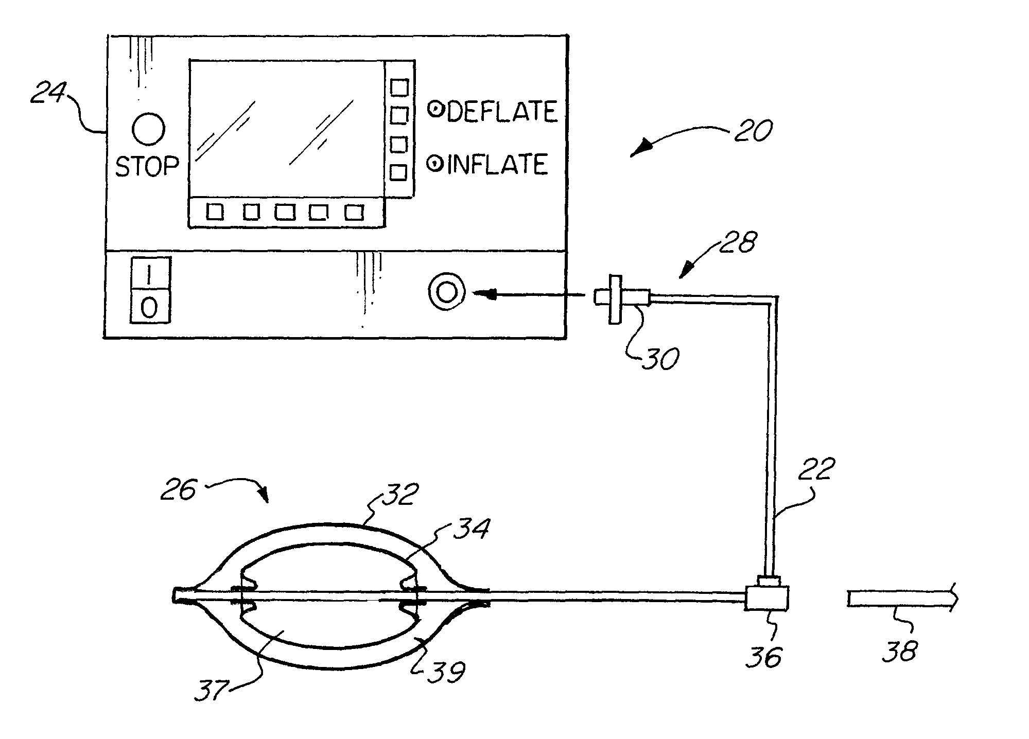
[0053]The basic components of one embodiment of a nested balloon catheter system in accordance with the invention are illustrated in FIG. 1. As used in the description, the terms “top,”“bottom,”“above,”“below,”“over,”“under,”“above,”“beneath,”“on top,”“underneath,”“up,”“down,”“upper,”“lower,”“front,”“rear,”“back,”“forward” and “backward” refer to the objects referenced when in the orientation illustrated in the drawings, which orientation is not necessary for achieving the objects of the invention.

[0054]As shown in FIG. 1, the nested balloon catheter system (20) includes a catheter (22) and a fluid source (24). The catheter (22) may have any suitable diameter and length depending on a particular application, and may be flexible, rigid or semi rigid. The catheter (22) may be made with any commercially available material that is flexible enough to allow the shaft to be safely inserted through the available opening of a bodily cavity such that it will bend instead of puncturing the wal...


PUM


Abstract

A balloon catheter for delivering a therapeutic and / or diagnostic agent to tissue is described including an outer balloon having a wall with an opening therethrough and an inner surface, an inner balloon disposed in the outer balloon, enclosing an inflation chamber and having an outer surface defining a space between the outer surface of the inner balloon and the inner surface of the outer balloon, a catheter having a first lumen in fluid communication with the space between the inner balloon and the outer balloon for supplying the agent thereto, and a second lumen through which fluid is supplied to the inflation chamber for inflating the inner balloon to urge the agent out of the opening in the outer balloon, wherein the outer balloon and / or the inner balloon comprise at least one protrusion for directing the agent.

Description

FIELD OF THE INVENTION[0001]The present invention relates to methods and systems for delivering therapeutic and / or diagnostic agents to specific cellular locations within and adjacent to bodily tissues and cavities. More specifically, the invention relates to a method and system of localized delivery of diagnostic and / or therapeutic agents to bodily tissues and cavities via a nested balloon catheter.BACKGROUND OF THE INVENTION[0002]In diagnosing and treating diseases of various body cavities and organs, it is necessary to deliver diagnostic and / or therapeutic agents to the organs at specified locations. Most common routes of drug delivery include a non-invasive peroral (through the mouth), topical (skin), transmucosal (nasal, buccal / sublingual, vaginal, ocular and rectal) and inhalation routes. However, many therapeutic and diagnostic agents in general may not be delivered using these routes because they might be susceptible to enzymatic degradation or cannot be absorbed into the sy...

Claims


Application Information

Patent Timeline
Patent Type & Authority Patents(United States)
IPC IPC(8): A61M25/10A61F2/958
CPCA61M25/1011A61M5/007A61B5/065A61M25/1018A61M2025/1013A61M2025/105A61M2025/1052A61M2025/1086A61N5/0603A61N5/1014A61B5/021A61B5/4839A61M25/10184
Inventor GERRANS, LAWRENCE J.GUNDAY, ERHAN H.
Owner SANOVAS
Why Eureka
Try Eureka