Multimodal imaging of atherosclerotic plaque targeted to LOX-1
a multi-modal imaging and atherosclerotic technology, applied in the direction of angiography, drug composition, dispersed delivery, etc., can solve the problem of no clinically established non-invasive method for staging atherosclerotic lesions
- Summary
- Abstract
- Description
- Claims
- Application Information
AI Technical Summary
Benefits of technology
Problems solved by technology
Method used
Image

Examples
embodiments
[0044]Many studies have demonstrated that oxidized low density lipoprotein (ox-LDL) plays a critical role in atherogenesis. The ox-LDL receptor LOX-1 mediates the pathological effects of ox-LDL in atherosclerotic lesions. LOX-1 is a type II transmembrane protein with a short intracellular cytoplasmic tail and a long extracellular domain containing a C-type lectin-like structure. LOX-1 induces free radical generation, apoptosis of endothelial cells and monocytes / macrophages, expression of adhesion molecules and activates the inflammatory cascade. LOX-1 also induces expression and activation of matrix metalloproteinases (MMPs). These pathological effects of LOX-1 not only initiate atherosclerotic lesion formation, but also contribute to the vulnerability of a plaque to rupture. A recent study (Mehta et al., Circulation Research, 2007; 100:1634-1642) demonstrated that LOX-1 deficiency significantly decreases the formation of atherosclerotic lesions. In addition, LOX-1 is highly express...
example 1
Cardiovascular Magnetic Resonance (CMR) Based Molecular Imaging of LOX-1
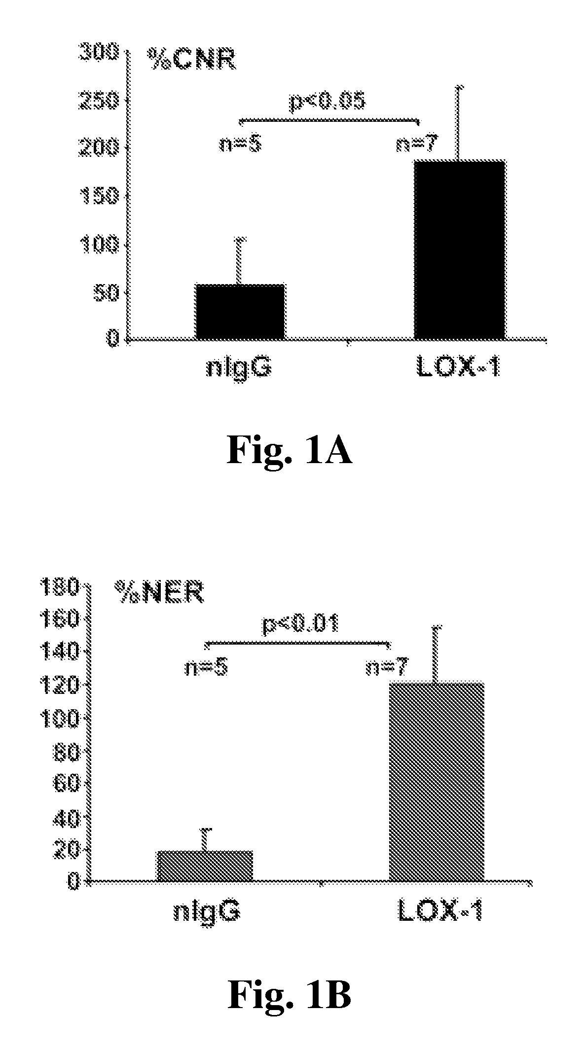
[0067]Oxidized low-density lipoprotein and its receptor LOX-1 play a crucial role in the initiation, progression, and destabilization of atherosclerotic lesions. A noninvasive tool to improve the clinical characterization of this pathological process is needed. The following study indicates the feasibility of CMR based molecular imaging compositions targeted to LOX-1 which is highly expressed on atherosclerotic lesions in mice.
[0068]Materials and Methods:
[0069]LDLR− / − mice on an atherogenic diet for >16 weeks were used. The imaging probe consisted of liposomes decorated with anti-LOX-1 antibody (or nonspecific IgG), gadolinium, and DiI fluorescence markers. MRI at 7.0 T (Clinscan, Bruker / Siemens) was performed at baseline and 24 hrs after intravenous injection of 150 ml of probe containing LOX-1 antibody (n=7) or nonspecific IgG (nIgG) (n=5) with 0.075 mmol Gd / kg, followed by excision of the aorta for frozen cro...
example 2
Radiolabel Based Molecular Imaging of LOX-1
[0075]In the following experiment a novel radio labeled imaging probe targeted to LOX-1 was used to assess the presence of atherosclerosis plaques.
[0076]Materials and Methods:
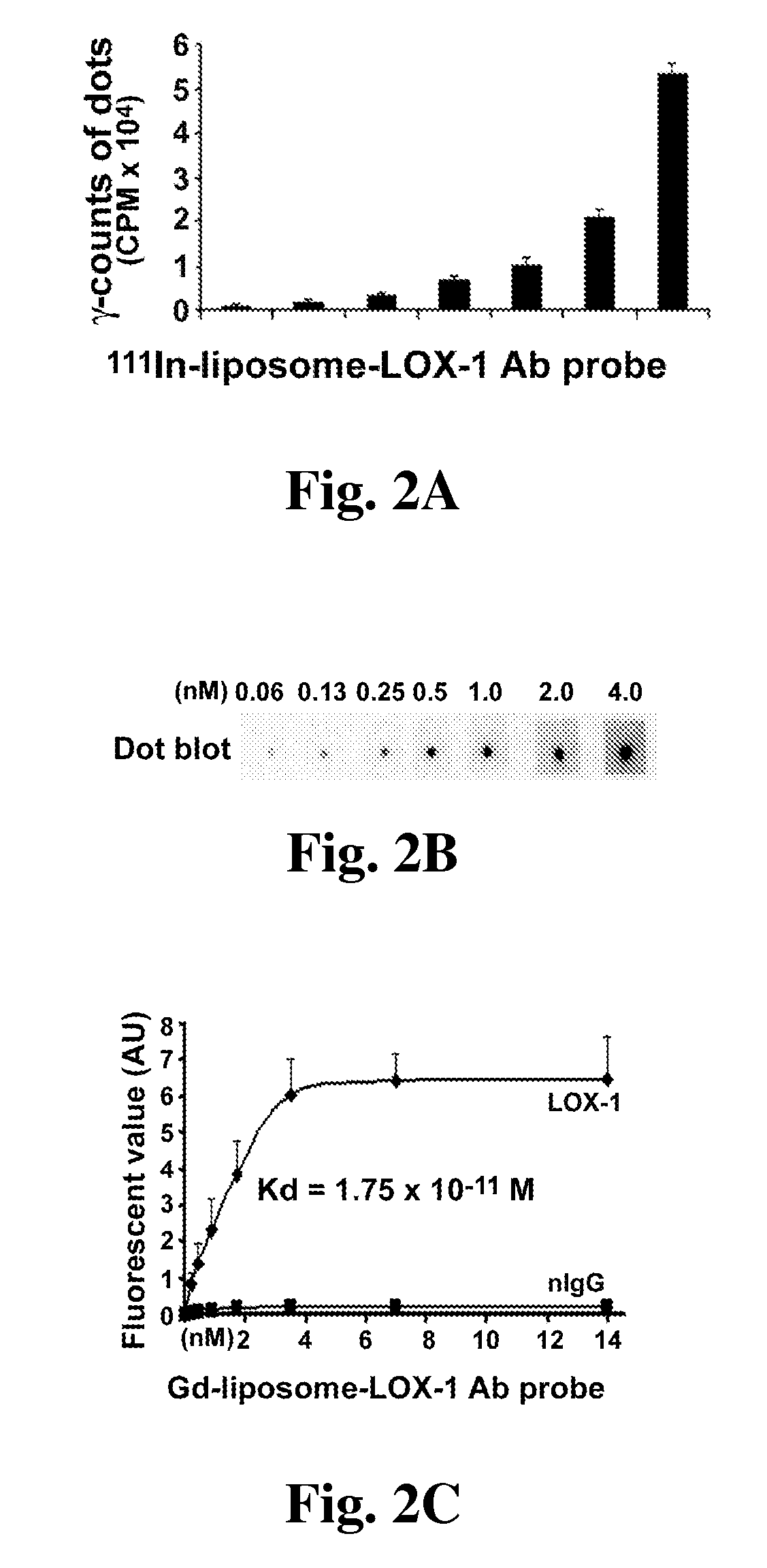
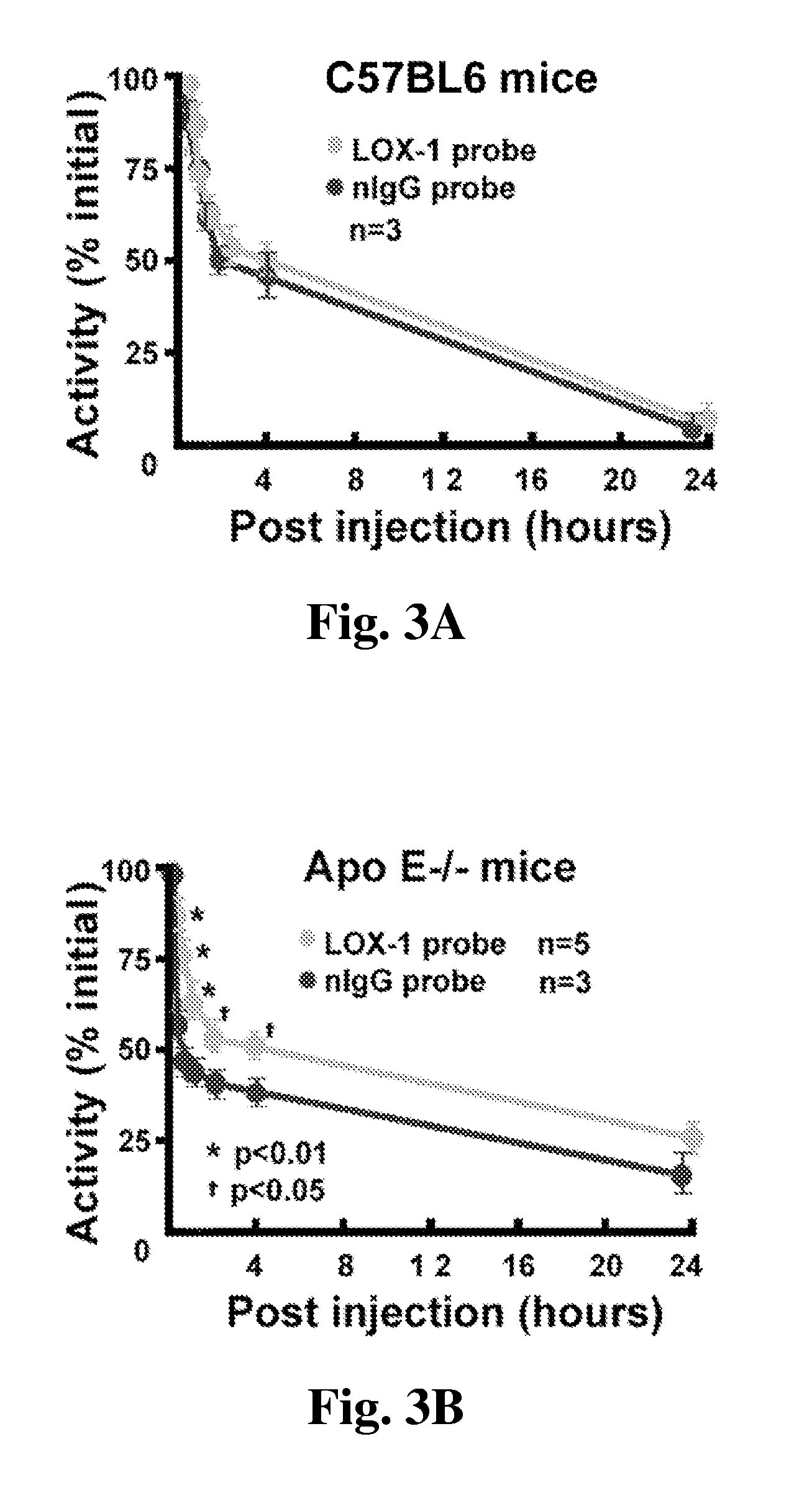
[0077]20 Apo E− / − mice on Western diet for >20 weeks were used for this experiment. The targeted imaging probe (TP) consisted of liposomes decorated with anti-LOX-1 antibody, 111In and DiI fluorescence markers. Control probe (CP) carried nonspecific IgG. 24 hrs after iv probe injection, 5 mice per group were used for ex vivo fluorescence microscopy. The other 5 mice were used for microSPECT / CT, followed by ex vivo aorta phosphor imaging.
[0078]Results:
[0079]The in vitro binding study demonstrated selective probe targeting to LOX-1. In vivo fluorescence imaging showed that the targeted imaging probe, but not control probe, bound mainly to the shoulder area of some plaques. The targeted imaging probe signal in plaques was co-localized with the areas of apoptosis and eleva...
PUM
| Property | Measurement | Unit |
|---|---|---|
| mean diameter | aaaaa | aaaaa |
| molecular weight | aaaaa | aaaaa |
| diameter | aaaaa | aaaaa |
Abstract
Description
Claims
Application Information