Method for determining locations of implanted electrodes with medical images

a technology of medical images and electrodes, applied in the field of medical imaging systems and methods, can solve the problems of inability to accurately predict the location of implanted electrodes, difficult to plan surgical margins for epileptic zone resection, and inaccurate cerebral models when ray tracing is implemented

Active Publication Date: 2014-03-04
THE MEDICAL COLLEGE OF WISCONSIN INC
View PDF23 Cites 8 Cited by
  • Summary
  • Abstract
  • Description
  • Claims
  • Application Information

AI Technical Summary

Benefits of technology

[0011]The present invention overcomes the aforementioned drawbacks by providing a method for the registration of implanted electrode locations, such as implanted intracranial electroencephalography (“iEEG”) electrode locations, with a subject-specific 3D brain model produced from post-implantation magnetic resonance (“MR”) images of the subject. More particularly, a method is provided for the determination of the locations of such implanted intracranial electrodes. Subsequently, the locations are displayed on a three-dimensional rendering of a subject-specific, post-implantation cortical surface reconstruction. This rendering, or brain model, is produced using a method that begins with seed points located, for example, at the boundary between gray matter and white matter in 3D MR images, and then builds the model outward towards the cortical surface. This method utilizes the combination of post-implantation CT and MR imaging, and provides 3D representations of each subject's uniquely compressed, cortical surface anatomy with iEEG electrode locations overlaid. It also allows for midline cortical surface views of electrodes placed deep between the cerebral hemispheres in the interhemispheric fissure, previously unobtainable using whole-brain 3D volume rendering techniques. Additionally, a method is provided for creating illustrative electrode “shadows” that identify cortical patches beneath each electrode and help locate sulcal-straddling electrodes that potentially record iEEG signals from two adjacent gyri.
[0013]It is another aspect of the invention to provide a method for implanted electrode localization that requires substantially no manual steps in determining electrode position. Instead, electrode locations are physically measured in co-registered CT images and overlaid on a subject-specific 3D model of the brain. This automation minimizes human error in producing reliable images for use in, for example, epilepsy surgery planning.

Problems solved by technology

Planning surgical margins for resection of epileptic zones is a challenging problem due to the uncertainty associated with subdural electrode positions and inaccurate, labor-intensive techniques for electrode localization.
The noted limitation of such methods is that susceptibility artifacts resulting from the presence of implanted electrodes are pervasive in MR images, thereby yielding inaccurate cerebral models when ray tracing is implemented.
These methods are useful in the visualization of grid and strip locations, but lack a degree of accuracy and / or resolution in imaging detail due to interpolation and projection limitations.
Many of these techniques also rely on expert guidance and manual marking of electrode positions, making them prone to human measurement error.
Thus many electrodes cannot be localized by intra-operative photography, especially strip electrodes placed via small burr hole craniotomies.
X-ray image projection corrects for this brain distortion when x-ray image acquisition follows the closure of the craniotomy defect; however, 2D x-ray images still require elaborate registration techniques for 3D visualization on cortical surfaces, such as those described by S. S. Dalai, et al., in “Localization of Neurosurgically Implanted Electrodes Via Photograph-MRI-Radiograph Coregistration,”J. Neurosci. Methods, 2008; 174:106-115, and are not currently realizable without dedicated experts.
This method ultimately lacks 3D information required for visualizing foreshortened electrode strips oriented along the x-ray path rather than perpendicular to the x-ray beam, and lacks information for medially located electrodes.
This is done because MRI images gathered post-implantation are subject to magnetic field susceptibility artifacts caused by the metal electrodes at the surface of the brain.
3D rendering of electrode-affected images has thus been limited, and subsequently brain shift and compression caused by electrode grids and craniotomy defects have never been addressed in imaging models.
Despite the numerous and varied technical solutions developed over a wide span of years, as described above in detail, none have yet been able to provide such a method.

Method used


Image

  • Method for determining locations of implanted electrodes with medical images
  • Method for determining locations of implanted electrodes with medical images
  • Method for determining locations of implanted electrodes with medical images

Examples


Embodiment Construction

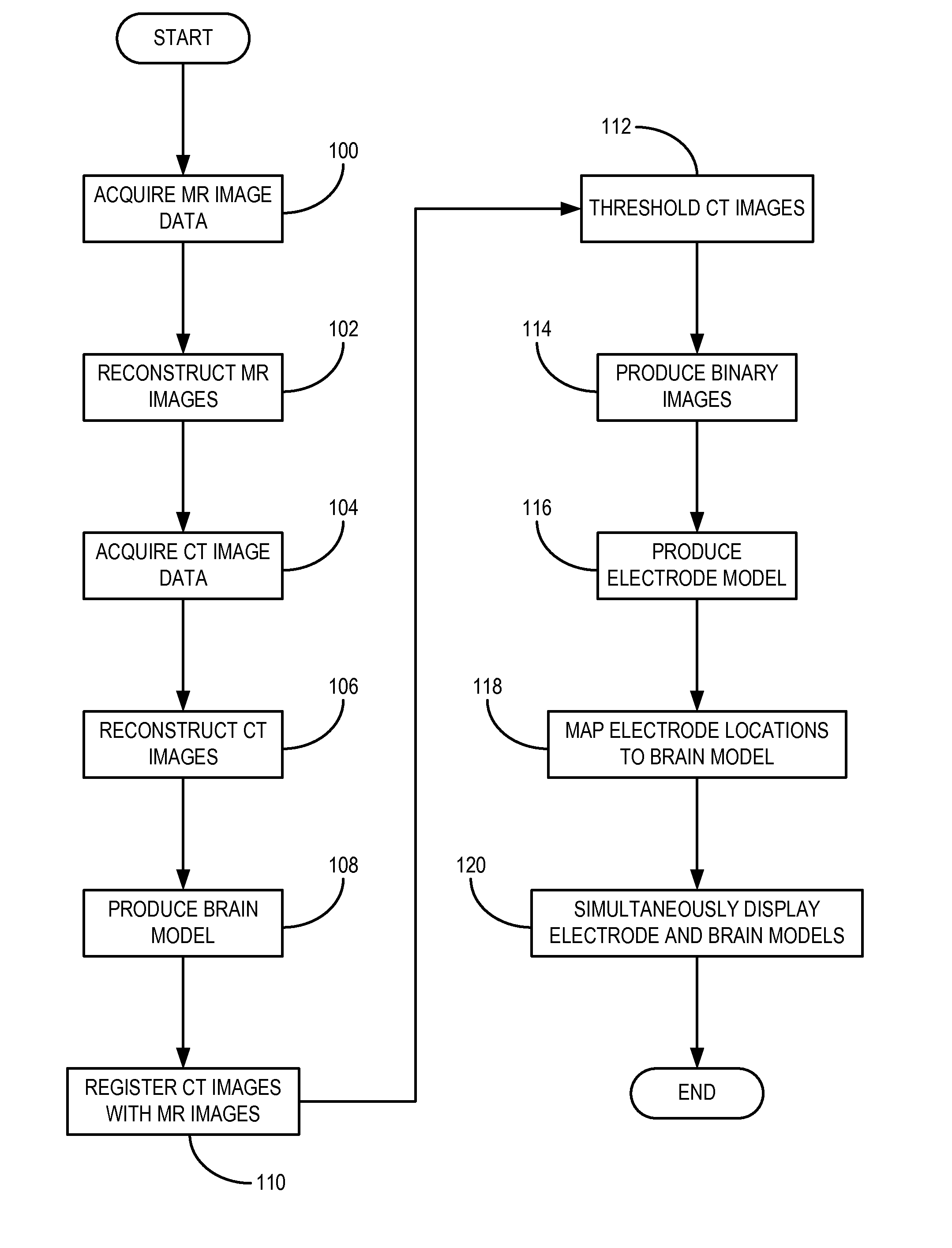
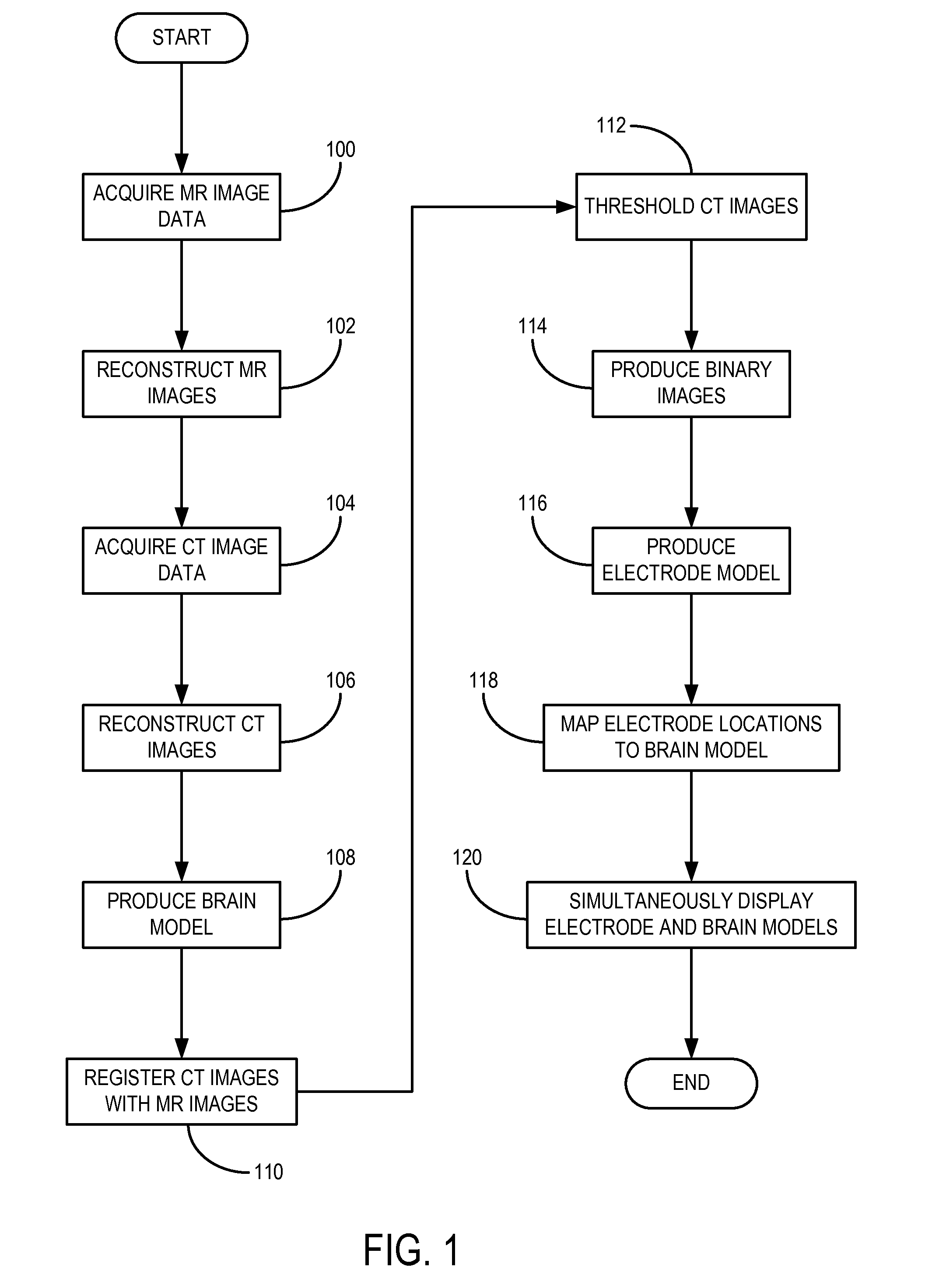
[0024]Surgical resection of cortical areas that initiate seizures can be an effective treatment option for patients with, for example, drug-resistant partial epilepsy. Intracranial electroencephalography (“iEEG”) studies are often necessary for localizing seizure onset zones. These multichannel EEG tracings are traditionally analyzed visually to identify surgical targets. To acquire these tracing, subdural, or intracranial, electrodes are surgically implanted in the subject's brain. Exemplary electrodes include platinum-iridium alloy electrode discs (Ad-Tech Medical, Racine, Wis.) having a diameter, for example, of 4 millimeters (“mm”). Such exemplary electrodes are arranged in any number of particular arrangements, including, for example, a grid (e.g., 8×8), a partial grid (e.g., 8×2), and a strip (e.g., 4×1, 6×1, and 8×1). It should be appreciated by those skilled in the art that combinations of each of the preceding electrode arrangements are possible. Burr hole, or “trephine,” c...


PUM


Abstract

A method for accurate localization and visualization of implanted electrodes, such as implanted intracranial electrodes, is provided. More particularly, a realistic representation of intracranial electrode positions on patient-specific post-implantation MRI brain renderings is obtained. The resulting computer models provide an accurate depiction of electrode locations on three-dimensional brain renderings that are suitable for use in surgical planning of resection boundaries around, for example, epileptic zones. Electrodes placed inter-hemispherically are also visible with this method. In addition, a method for creating electrode “shadows” cast upon the brain model surface is provided. These electrode shadows are useful for estimating cortical areas sampled by iEEG and for locating electrodes that may straddle sulci and contact two adjacent cortical gyri.

Description

CROSS-REFERENCE TO RELATED APPLICATIONS[0001]This application represents the U.S. National Stage of International Application No. PCT / US2010 / 051946, filed Oct. 8, 2010 which claims the benefit of U.S. Provisional Patent Application Ser. No. 61 / 249,660 filed on Oct. 8, 2009, and entitled “Method for Determining Locations of Intracranial Electroencephalography Electrodes with Medical Images.”The foregoing applications are incorporated herein by reference in their-entirety.FIELD OF THE INVENTION[0002]The field of the invention is medical imaging systems and methods. More particularly, the invention relates to determining and visualizing the locations of implanted intracranial electrodes using a first set of post-implantation images, for example, acquired using magnetic resonance imaging (“MRI”) and a second set of post-implantation images, for example, images acquired using x-ray computed tomography (“CT”).BACKGROUND OF THE INVENTION[0003]Subdural electrodes are placed on the brains of...

Claims


Application Information

Patent Timeline
Patent Type & Authority Patents(United States)
IPC IPC(8): A61B5/05
CPCA61B6/501A61B6/12A61B5/0478A61B5/06A61B6/037A61B6/032A61B6/5247A61B5/4094A61B5/291A61B5/293
Inventor LAVIOLETTE, PETER S.RAND, SCOTT D.
Owner THE MEDICAL COLLEGE OF WISCONSIN INC
Why Eureka
Try Eureka