Peptide inhibition of lung epithelial apoptosis and pulmonary fibrosis

a technology of pulmonary fibrosis and apoptosis, which is applied in the field of biochemistry and medicine, can solve the problems of affecting the pathogenesis of lung injury and pulmonary fibrosis, limiting the understanding of the regulatory interactions of these molecules, and affecting the survival of lung cells, so as to enhance the expression of pro-survival (or anti-apoptotic) molecules, block bleo-induced mouse lung fibrosis, and improve the effect of l

Active Publication Date: 2014-04-15
BOARD OF RGT THE UNIV OF TEXAS SYST
View PDF6 Cites 9 Cited by
  • Summary
  • Abstract
  • Description
  • Claims
  • Application Information

AI Technical Summary

Benefits of technology

[0059]The present inventors have discovered that treatment with either of two peptides, PP-1 and PP-2 (sequences provided below), that are believed to act by inhibition of uPAR-Cav-1 interactions and PAR-β1-integrin associated Src kinase activity, protect LECs from bleo-induced apoptosis. This is believed to be mediated by suppression of the proapototic molecules p53 and PAI-1 while enhancing the expression of pro-survival (or anti-apoptotic) molecules uPA and uPAR.

Problems solved by technology

While these interactions comprise a new paradigm, their role in the pathogenesis of lung injury and pulmonary fibrosis is not yet fully elucidated.
Understanding the regulatory interactions of these molecules is currently limited.
Complete deficiency of p53 would be deleterious, permitting the persistence of severely damaged LECs and other damaged lung resident cells.
CSD targeted GFP to caveolae, albeit less efficiently than full-length Cav-1
However, it was noted that the physiological importance of Cav-1 in regulating signaling has been difficult to distinguish from its traditional functions in caveolar assembly, transcytosis, and cholesterol transport.
The authors concluded that Cav-1, may interfere with synaptic facilitation and long term potentiation formation in the hippocampus.

Method used


Image

  • Peptide inhibition of lung epithelial apoptosis and pulmonary fibrosis
  • Peptide inhibition of lung epithelial apoptosis and pulmonary fibrosis
  • Peptide inhibition of lung epithelial apoptosis and pulmonary fibrosis

Examples


example i

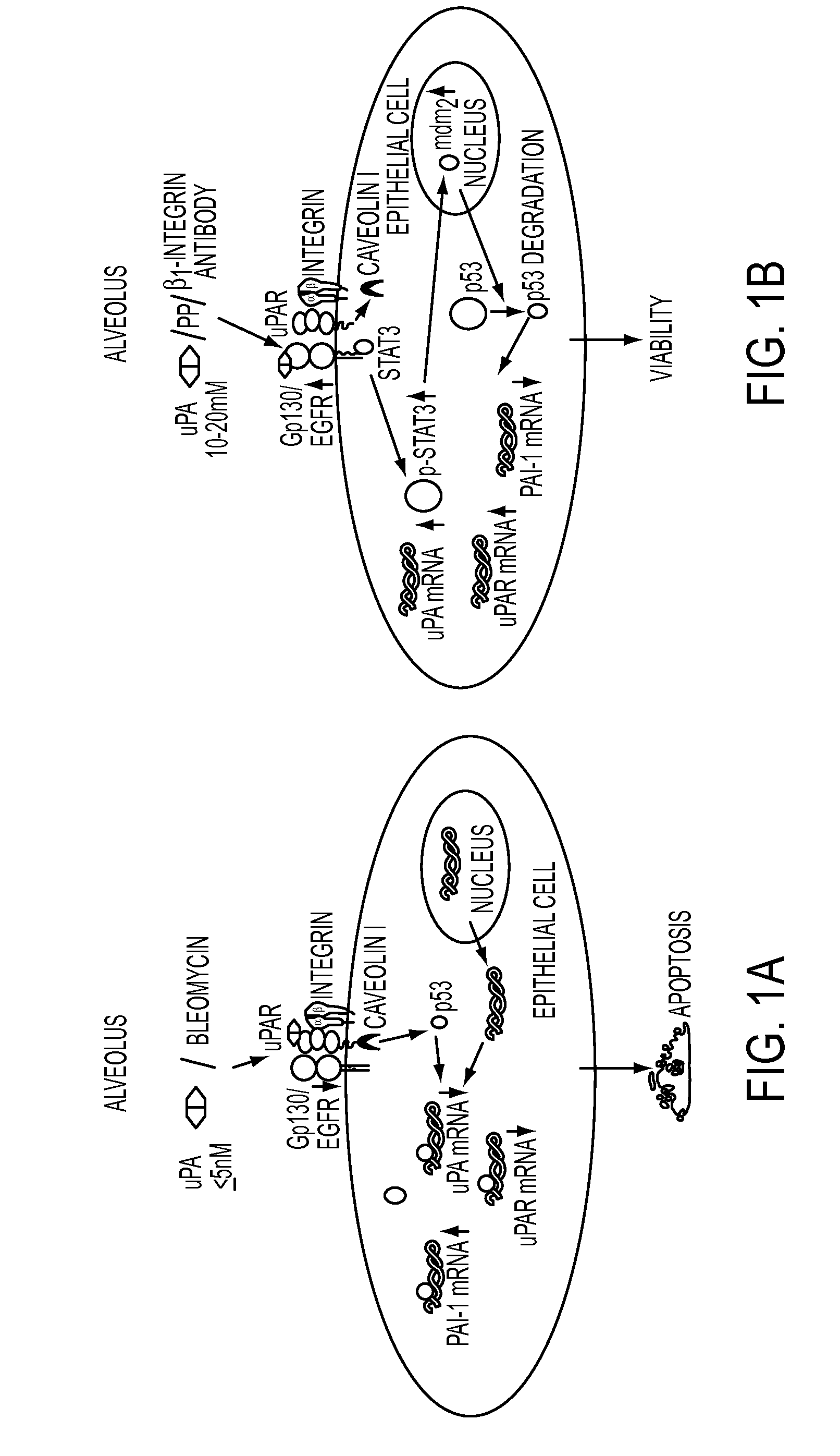
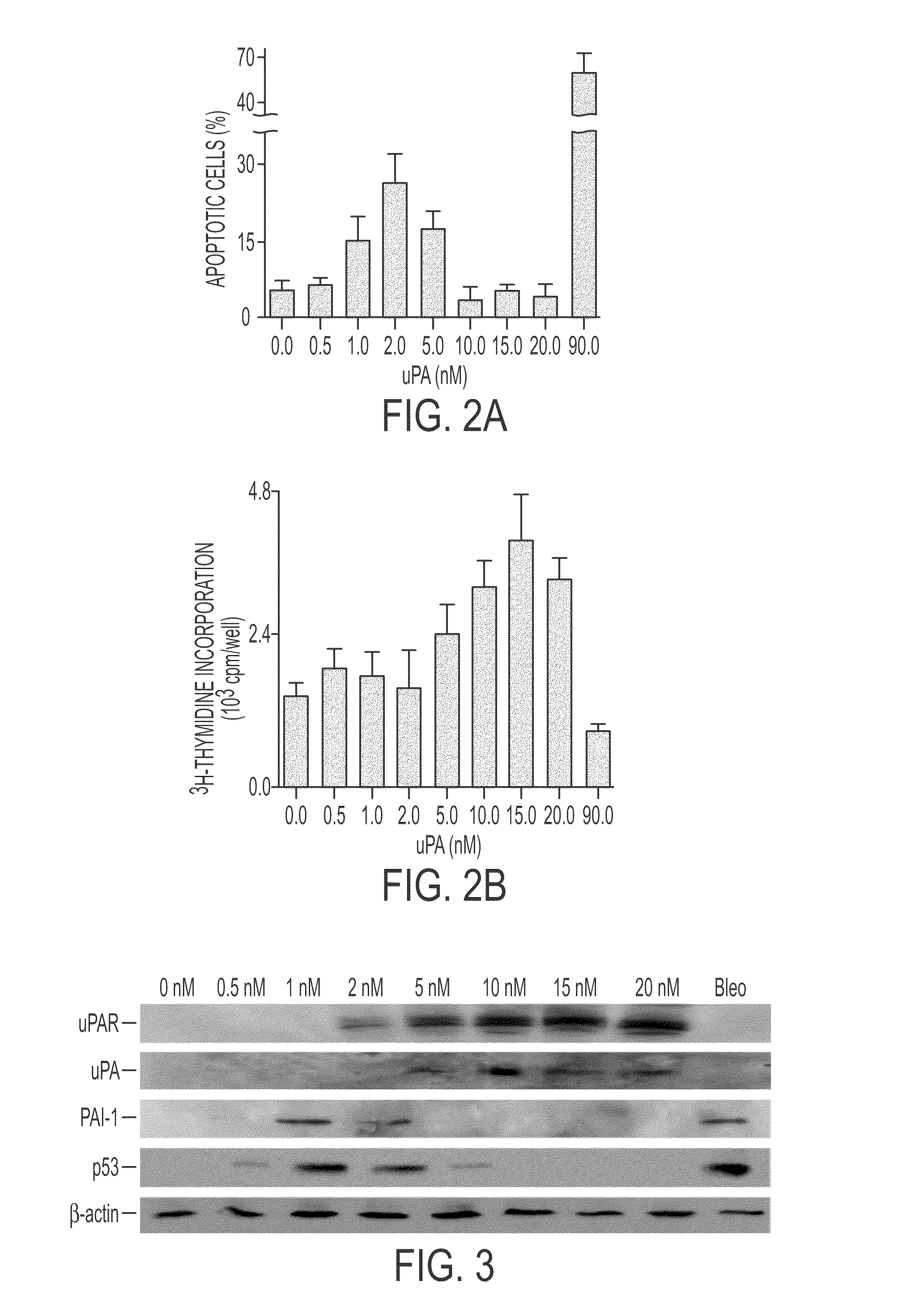
uPA Induces Dose-Related Apoptosis and Proliferation of Lung Epithelial Cells

[0234]uPA treatment of primary small airway epithelial (SAE) cells in vitro for 48 hours induced apoptosis with an inverted U-shaped dose-response curve over a range of 0.5-20 nM. Cell proliferation is also induced by concentrations of between 5 and 20 nM. The apoptotic effects were weaker than those of bleomycin which did not induced, but rather inhibited cell proliferation. These results appear in FIGS. 2A-2B.

[0235]Western blots were made of extracts of uPA treated cells obtained 24 hours after the uPA was added. Results in FIG. 3 show that both uPA and uPAR expression was induced whereas PAI-1 and p53 expression was suppressed.

example ii

The Effect of Bleo on LEC uPA, uPAR, PAI-1 and p53 Expression In Vitro

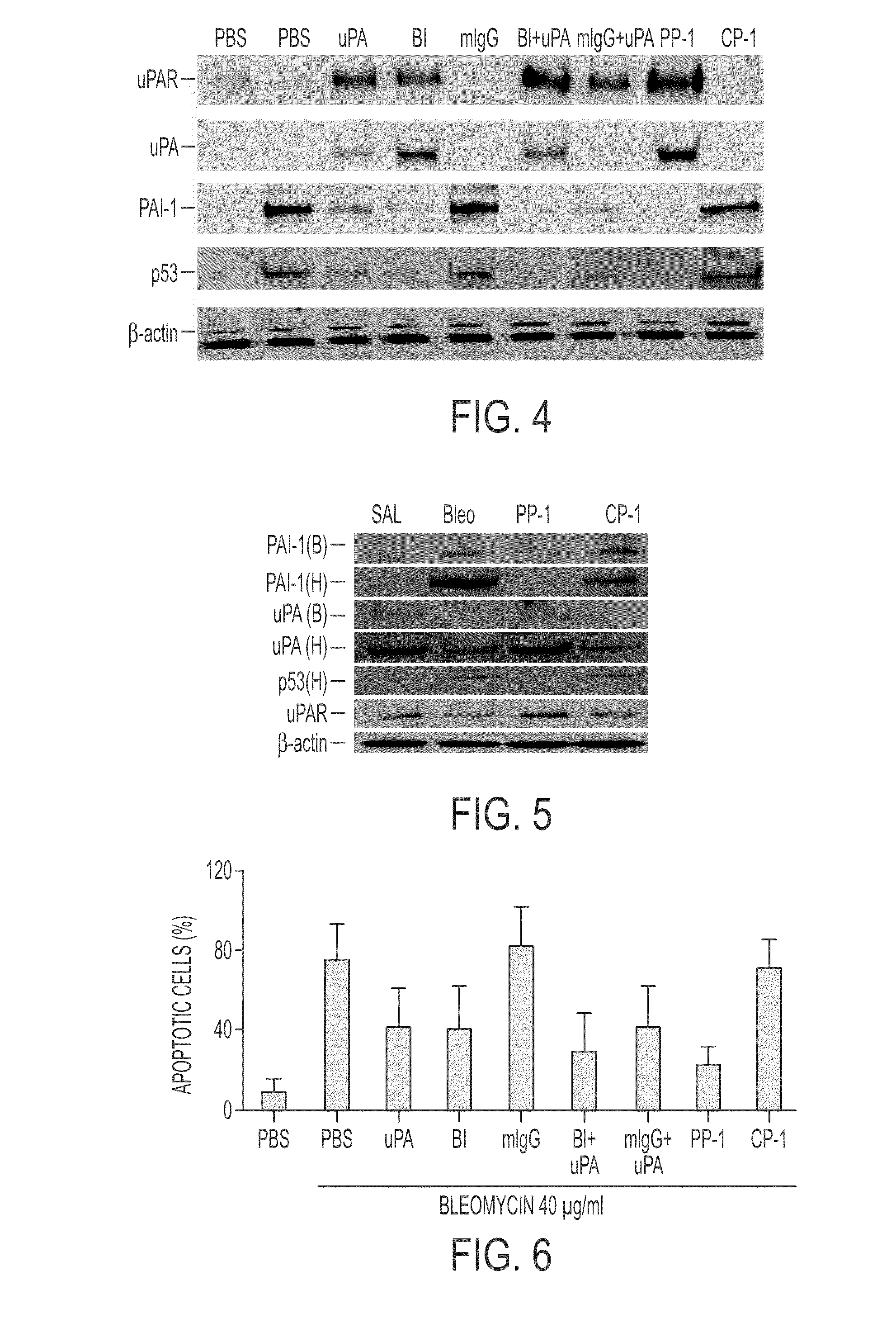
[0236]Beas2B cells were treated for 28 h with PBS or 40 μg / ml of bleo in the presence or absence of uPA or its ATF (20 nM), anti-β1-integrin monoclonal antibody (B1), (which activates β1-integrin by clustering), mouse IgG (mIgG), uPA+B1, uPA+mIgG, PP-1 (10 nM), or scrambled control peptide CP-1 (10 nM) at 37° C. In the case of uPA and PAI-1, the full length uPA was substituted with ATF, because uPA-mediated autoinduction or induction of PAI-1 occurs via the interaction of the ATF with uPAR (3-4). The cell lysates (CL) were analyzed for uPA, PAI-1, p53 and β-actin expression by Western blotting using specific antibodies. The membrane proteins were subjected to Western blotting using anti-uPAR antibody. The same responses of PAI-1 and uPA were seen in the conditioned medium (CM) by Western blotting (not shown). Alternatively, immunoprecipitation of cells labeled with 35S-methionine showed the same patterns of protei...

example iii

Induction or Suppression of Lung or BAL Protein Expression by Bleomycin and Effects of PP-1

[0238]Mice were treated intranasally with 50 μl saline or an equal volume of a 40 μg bleomycin solution. After 24 h, 10 mM PP-1 or the same concentration of control peptide CP-1 were given intraperitoneally using a 0.1 ml osmotic pump. Lungs were excised on day 7.

[0239]Lung tissue was chopped into small pieces with fine scissors and rinsed 3× with PBS. The tissues were then homogenized in 1 ml of extraction buffer (25 mM Tris-HCl, pH 7.9. 0.5 mM EDTA and 0.1 mM PMSF) and the homogenate was centrifuged at 12,000 g for 15 min at 4° C. The supernatant was removed and referred to as the crude extract. Protein content was measured using a BioRad protein assay kit. Cytosolic proteins were separated by SDS-PAGE and transferred to a nitrocellulose membrane which was blocked with 1% BSA in a wash buffer for 1 h at room temperature followed by overnight incubation with antibodies specific for uPA, PAI-1...


PUM

PropertyMeasurementUnit
molecular weightaaaaaaaaaa
timeaaaaaaaaaa
volumeaaaaaaaaaa

Abstract

During lung injury, p53 expression increases, inducing plasminogen activator inhibitor-1 (PAI-1) while inhibiting expression of urokinase-type plasminogen activator (uPA) and its receptor (uPAR), resulting in apoptosis of lung epithelial cells (LECs). In the bleomycin lung injury model, p53 and PAI-1 are induced while uPA and uPAR are inhibited. A 20 residue peptide DGIWKASFTTFTVTKYWFYR termed PP-1 (the Cav-1 scaffolding domain) or peptide NYHYLESSMTALYTLGH, termed PP-2, protected LECs from bleomycin-induced apoptosis in vitro and in vivo and prevented subsequent pulmonary fibrosis by attenuating lung epitheilial damage. Pharmaceutical compositions, peptide multimers and deliverable polypeptides comprising the above peptides are dislcosed. The peptides and functional variants, peptide multimers, cell-targeted polyepeptides and pharmaceutical compositions are used in methods for inhibiting apoptosis of injured or damaged lung epithelial cells and for treating acute lung injury and consequent pulmonary fibrosis.

Description

[0001]This application claims priority to provisional application 61 / 033,999, filed Mar. 5, 2008.STATEMENT OF RIGHTS TO INVENTIONS MADE UNDER FEDERALLY SPONSORED RESEARCH[0002]This invention was funded in part by a grant from the National Heart, Lung and Blood Institute, National Institutes of Health (R01-HL71147), which provides to the United States government certain rights in this invention.BACKGROUND OF THE INVENTION[0003]1. Field of the Invention[0004]The present invention in the field of biochemistry and medicine is directed to peptides designated PP-1 and PP-2, and functional variants thereof, that prevent apoptosis of lung epithelial cells in vitro and in vivo, and prevent or treat pulmonary fibrosis, making them useful for treating acute lung inflammation and its sequelae, primarily pulmonary fibrosis.[0005]2. Description of the Background Art[0006]Throughout the disclosure, references are cited either by a number(s) in parentheses, referring to the Documents list at the en...

Claims


Application Information

Patent Timeline
Patent Type & Authority Patents(United States)
IPC IPC(8): C07K11/00
CPCC07K14/705C07K2319/01
Inventor SHETTY, SREERAMAIDELL, STEVEN
Owner BOARD OF RGT THE UNIV OF TEXAS SYST
Why Eureka
Try Eureka