Endovascular device with membrane

a technology of endothelial cells and endothelial cells, which is applied in the field of endothelial cells, can solve the problems of restricted blood flow, ischemia or necrosis, occlusion of coronary arteries, etc., and achieve the effects of promoting thrombosis, enhancing endothelial cell migration and tissue in-growth, and reducing blood flow

Active Publication Date: 2014-05-06
MERLIN MD PTE
View PDF230 Cites 14 Cited by
  • Summary
  • Abstract
  • Description
  • Claims
  • Application Information

AI Technical Summary

Benefits of technology

[0018]In some embodiments, the porosity of the porous section is selected such that it enables enhanced endothelial cell migration and tissue in-growth for endothelialization while substantially preventing blood circulation to the diseased, damaged or weakened portion of the vessel wall.
[0066]In some embodiments, a method of treating a body vessel having an aneurysmal portion comprises the steps of: providing an endovascular device, comprising: an expandable member, expandable from a first position to a second position, said expandable member being expandable radially outwardly to the second position such that an outer surface of said expandable member engages with an inner surface of the body vessel so as to maintain a fluid pathway in said body vessel through a lumen in the expandable member; a membrane covering at least a portion of an outer surface of said expandable member; a plurality of pores in a porous section of the membrane, the porous section having a substantially uniform porosity over a length extending from a proximal end to a distal end of the porous section, porosity being determined by a pore spacing and a pore size; wherein the proportion of the total area of an outer surface of the porous section that consists of membrane material defines a material ratio; wherein the substantially uniform porosity is selected such that, when the expandable member is positioned in the body vessel, the membrane permits a flow of blood from within the lumen of the expandable member, through at least one of the pores, and into at least one branch vessel that branches off of the body vessel; and wherein the substantially uniform porosity is further selected such that, when the expandable member is positioned in the body vessel, the membrane reduces blood flow to the aneurysmal portion of the body vessel, promoting thrombosis at or in the aneurysmal portion; and positioning the expandable member in the body vessel.
[0067]In some embodiments, the porosity of the membrane is selected such that it enhances endothelial cell migration and tissue in-growth.
[0079]In some embodiments, the method further comprises providing a membrane having at least one surface-modifying end group that encourages healing of the body vessel after the device is inserted.

Problems solved by technology

Vessel occlusion or rupture of an aneurysm within the brain can result in stroke.
Aneurysms fed by intracranial arteries can grow within the brain to a point where their size can also cause a stroke or the symptoms of a stroke, requiring surgery to remove the aneurysm, or other remedial intervention.
Occlusion of coronary arteries is a common cause of heart attack.
Diseased and obstructed coronary arteries result in restricted blood flow in the heart which can lead to ischemia or necrosis.
Conventional open-heart surgery is of course highly invasive and traumatic for patients undergoing such procedures.
In both methods, a certain incidence of restenosis (resealing) occurs resulting in a loss of the benefit of the procedure, and potentially the need for additional rounds of therapy.
Restenosis also results in reversion back to the original occluded condition, such that the vessel no longer conducts a normal flow volume, which can lead to ischemia or infarct depending on the particular location and function of the vessel in question.
Foreign bodies frequently cause an inflammatory response, and in the case of blood vessels, neointimal proliferation which results in narrowing and occlusion of the body vessel, obviating the benefit of the implant.
For example, polyurethanes have been used in long term implants, but are not always suitable for use in endovascular treatments, especially in small blood vessels.
In addition, many commercially available polymers are with additives, or have impurities, that are surface-active and so reduce their usefulness in some biological applications.
However, each of these methods suffers from one or more problems including poor control of release or limitations of the form of drug or other reagent that can be applied.
Also, these methods are unsuitable for situations where it would be desirable to maintain the bioactive molecule on the device rather than having it be released, in order to maintain a relatively high local activity of the reagent of interest.
First, the stent must adequately support the diseased or weakened region in order to prevent rupture of the aneurysm or vessel, either of which could lead to serious complications including death, depending on the size, location and nature of the aneurysm or defect.
Second, in the case of stents use in the repair of aneurysms, the stent must permit sufficient blood to maintain the patency of both the parent and perforator vessels, while at the same time limiting flow to the aneurysm proper.
However, in biological applications, regulating movement of materials on cellular or subcellular scale is required, and it is difficult and costly to manufacture stents that have such fine effective pore size.
However, a problem inherent with these designs is that they are not easily adapted for effecting vessel wall repairs where the area of disease, damage or weakness can vary in size.
Furthermore, these designs are not optimized such that they will generally provide flow to perforator vessels that are part of the collateral circulation in the area of the diseased, damaged, or weakened vessel, while blocking flow to an aneurysm.

Method used


Image

  • Endovascular device with membrane
  • Endovascular device with membrane
  • Endovascular device with membrane

Examples


Embodiment Construction

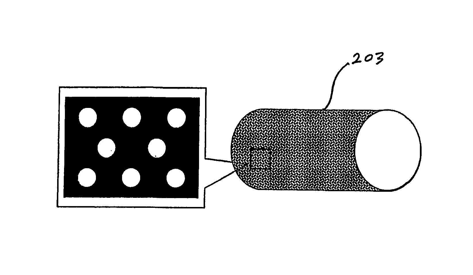
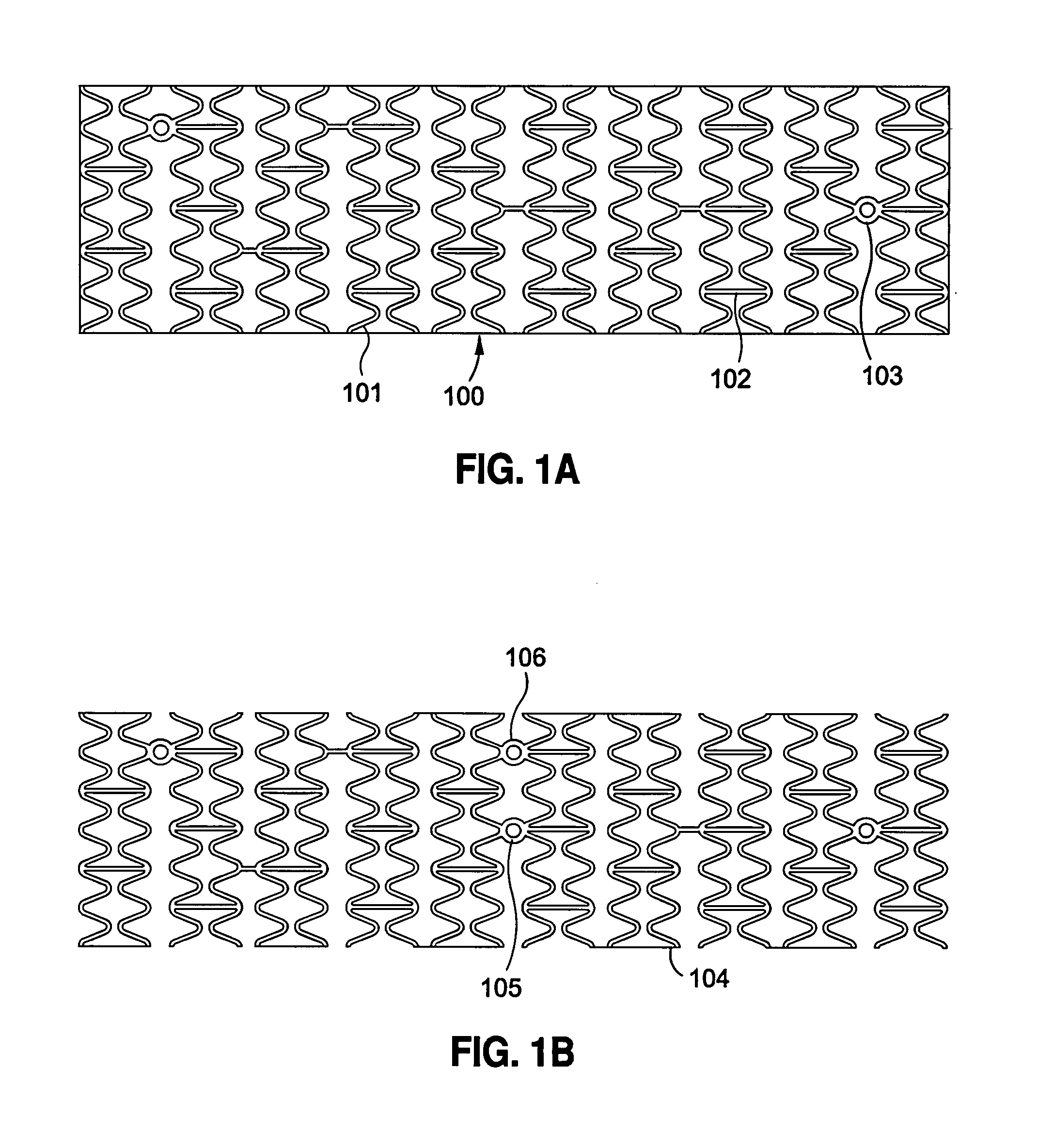
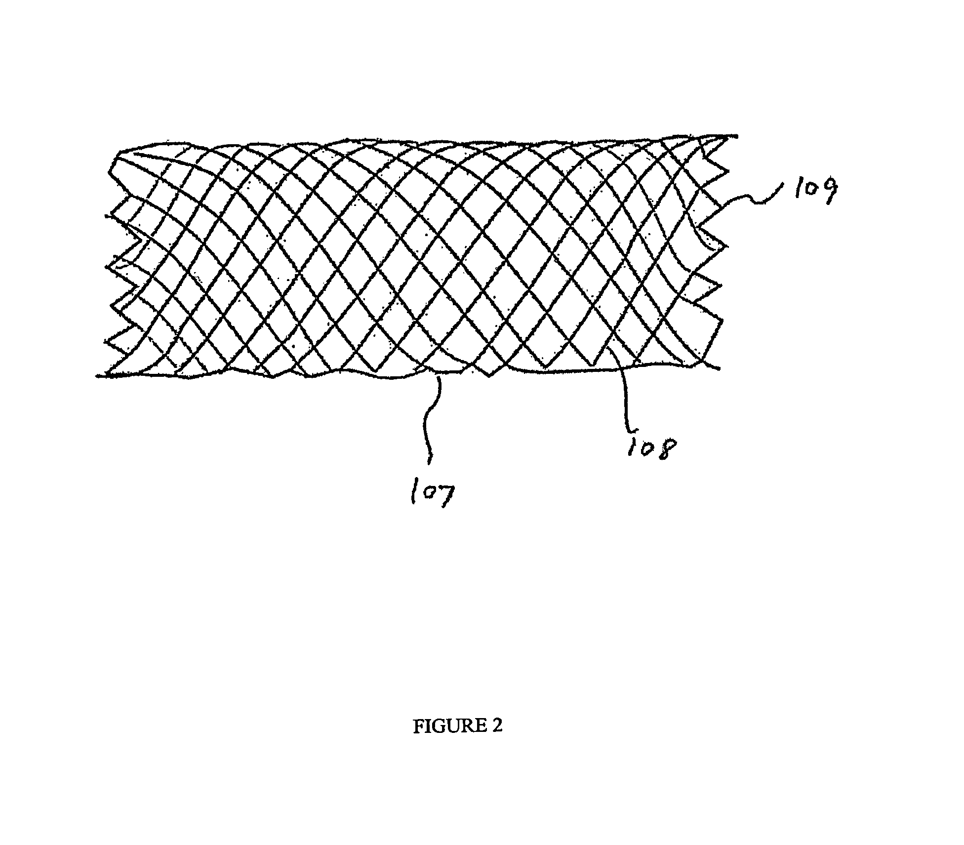
[0120]Implantable medical devices include physical structures for delivering drugs or reagents to desired sites within the endovascular system of a human body. These devices may take up diversified shapes and configurations depending upon specific applications. Common implantable medical devices include stents, vena cava filters, grafts and aneurysm coils.

[0121]The endovascular system of a human body includes blood vessels, cerebral circulation system, tracheo-bronchial system, the biliary hepatic system, the esophageal bowel system, and the urinary tract system. Although exemplary stents implantable in blood vessels are described, they are applicable to the remaining endovascular system. Embodiments of the invention, some of which are described herein are readily adaptable for use in the repair of a variety of vessels, including but not limited to, treatment or repair in cases of aneurysm, ischemic stroke, carotid artery stenosis, saphenous vein graft, small vessel stenosis, or ren...


PUM


Abstract

Embodiments of a uniformly porous membrane covering an endoprosthetic device, for example, a stent used to treat an aneurysm, are described. Some embodiments have a pore size and spacing that provides a material ratio of between 70-80% in the deployed state. Material ratio is the proportion of the total porous segment of the membrane that corresponds to membrane material, the remainder being pores. In some embodiments, pore size ranges from about 10-100 μm. In some embodiments, pores are equidistantly spaced with pore spacing in a range of about 40-100 μm. The combination of pore size and spacing are effective to provide a membrane that substantially prevents flow to the aneurysm, while maintaining flow to perforator vessels. In some embodiments the membrane includes permanently attached agents that promote attachment of endothelial cells or progenitors and healing of the aneurysm, or reduce immune responses detrimental to the healing process.

Description

RELATED APPLICATIONS[0001]The present application is a continuation-in-part of U.S. patent application Ser. No. 11 / 586,899, filed Oct. 25, 2006, which is a continuation-in-part of U.S. patent application Ser. No. 10 / 580,139, filed May 19, 2006, which is filed under 35 U.S.C. § 371 as a U.S. National Stage Application of PCT International Patent Application No. PCT / SG2004 / 000407, filed Dec. 13, 2004, which claims priority to Singapore Patent Application No. SG200401735-6, filed Mar. 31, 2004; the present application is also a continuation-in-part of U.S. patent application Ser.No. 11 / 637,188, filed Dec. 12, 2006, which claims priority to Singapore Patent Application SG200508026-2, filed Dec. 13, 2005; the present application is also a continuation-in-part of PCT International Patent Application No. PCT / SG2006 / 000028, filed Feb. 13, 2006; the contents of each of the aforementioned applications are hereby incorporated by reference herein in their entireties.FIELD OF THE INVENTION[0002]...

Claims


Application Information

Patent Timeline
Patent Type & Authority Patents(United States)
IPC IPC(8): A61F2/06
CPCA61F2/07A61F2/915A61F2002/065
Inventor RUDAKOV, LEONHONG, TSUI YING RACHELO'CONNOR, MICHAEL
Owner MERLIN MD PTE
Why Eureka
Try Eureka