Characteristic parameters of cells and tissue from quantitative phase imaging

a quantitative phase and cell technology, applied in the field of characteristic parameters of cells and tissues from quantitative phase imaging, can solve the problems of anisotropy factor g, direct measurement, and extremely difficult, and achieve the effect of reducing the number and improving the quality of spherical scattering parameters

Active Publication Date: 2015-01-06
THE BOARD OF TRUSTEES OF THE UNIV OF ILLINOIS
View PDF1 Cites 4 Cited by
  • Summary
  • Abstract
  • Description
  • Claims
  • Application Information

AI Technical Summary

Problems solved by technology

However, the direct measurement of these scattering parameters, which may include, but are not limited to, the scattering mean free path (MFP) ls and anisotropy factor g, is extremely challenging.In the absence of absorption, the scattering mean free path, ls, is the average distance between two adjacent scattering events or the distance over which the unscattered light decreases to 1 / e of its original power.
The direct measurement of the foregoing scattering parameters is extremely challenging and, therefore, simulations, such as Monte Carlo, or finite-difference time-domain simulations, are often used iteratively instead.
Despite these promising efforts, light scattering-based techniques currently have limited use in the clinic.
A great challenge is posed by the insufficient knowledge of the tissue optical properties.

Method used


Image

  • Characteristic parameters of cells and tissue from quantitative phase imaging
  • Characteristic parameters of cells and tissue from quantitative phase imaging
  • Characteristic parameters of cells and tissue from quantitative phase imaging

Examples


example i

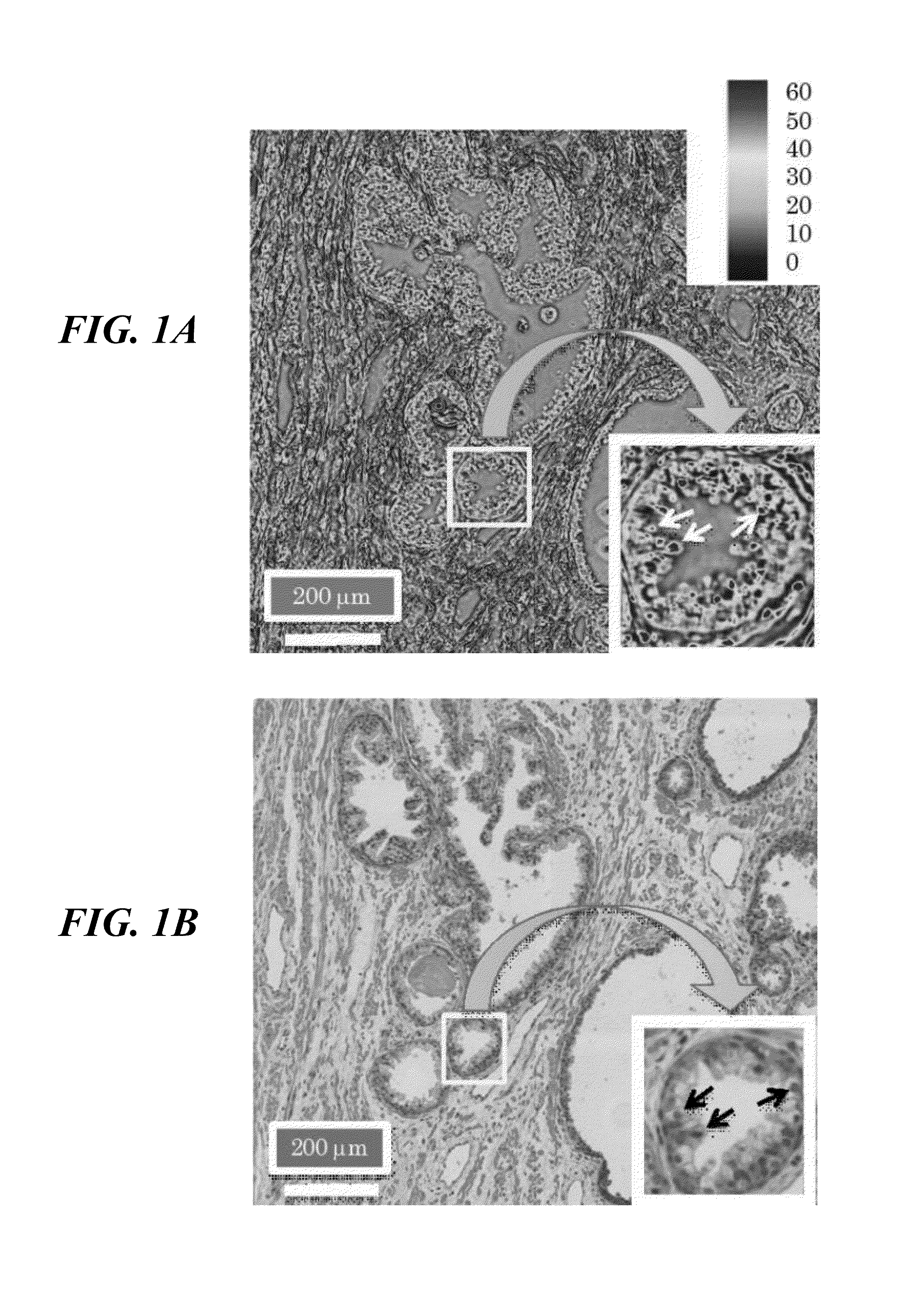
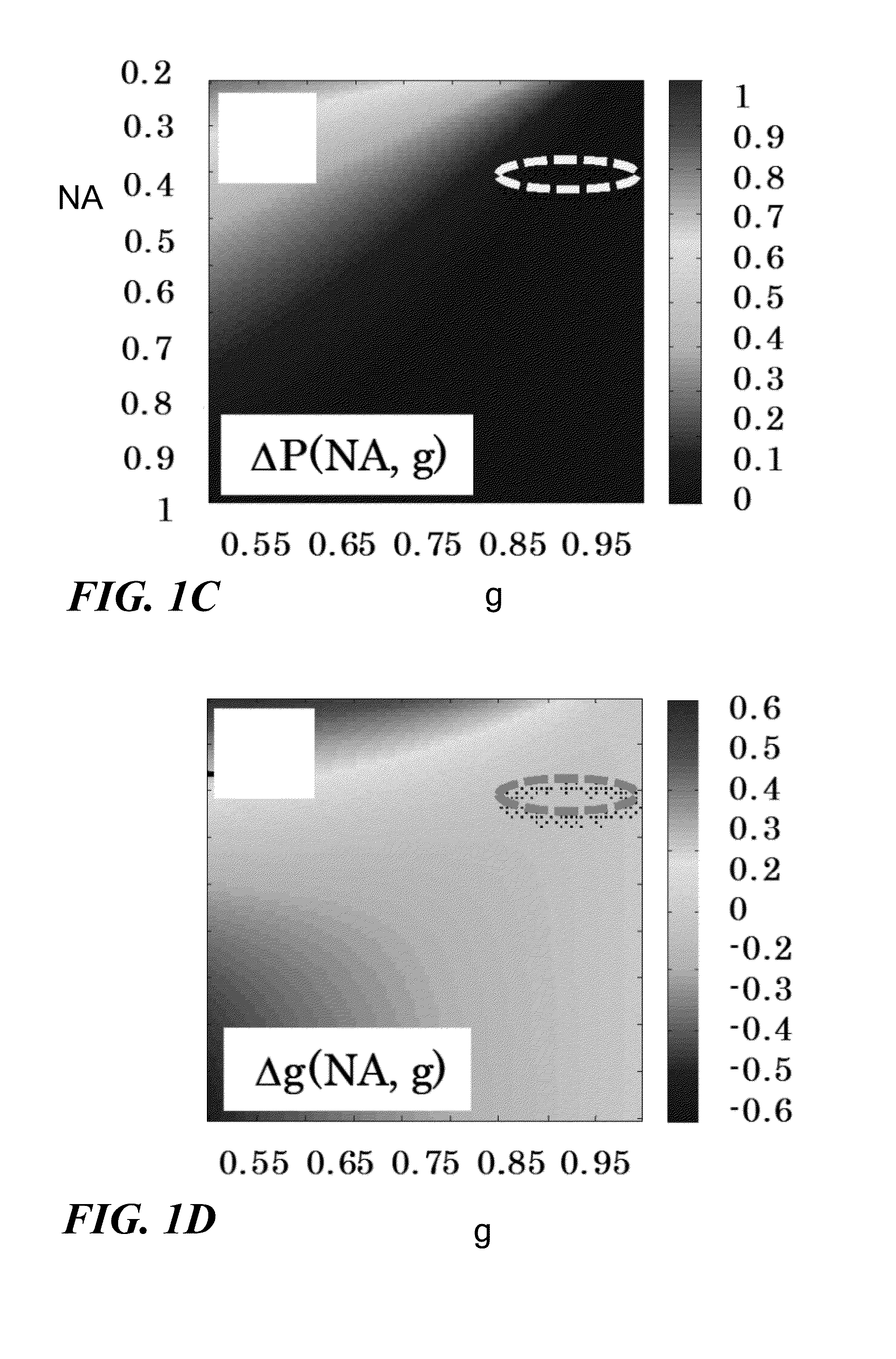
[0036]In an example of the application of the present invention, two adjacent 4 μm-thick tissue biopsies, one unstained and one stained by hematoxylin and eosin stain (H&E) were imaged by SLIM and in a bright field microscope, respectively. FIGS. 1A-1D illustrate the ability of SLIM to render high transverse resolution, high phase sensitivity images of thin tissue slices. The phase information provided by SLIM is inherently averaged over the optical frequencies, as discussed by Wang, et al., Appl. Phys. Lett., vol. 96, 051117 (2010), which is incorporated herein by reference. Thus, the scattering parameters obtained by this method will also be frequency-averaged. Throughout the experiments discussed here in the context of the present application, a 10×, 0.3 NA objective was used. This limited numerical aperture effectively acts as a low-pass spatial frequency filter.

[0037]The spatial averages performed in deriving equations 1a-b are expected to be affected by this cut-off of the lim...

example ii

[0041]In a further example of the application of the present invention, quantitative phase images associated with 5-1 μm thick tissue slices from rat organs were acquired. The tissue was sliced frozen but thawed before imaging. Three slices from each organ of the same rat were cut in succession and imaged by SLIM. The field of view of the microscope was 0.4×0.3 mm2. In order to image the cross-section of the entire organ, the specimen was translated and a mosaic of quantitative phase images was acquired and numerically collaged together. Single quantitative phase images made of hundreds of individual SLIM images were obtained. Note that these quantitative phase images cover the entire cross section of a rat organ, with a resolution of ˜λ / 2NA=0.9 μm. FIG. 2A shows one example of quantitative phase image of a tissue slice cut from a three month old rat liver.

[0042]Following Eq. 1, ls and g were calculated in windows of 9×9 μm2 across the entire tissue slice. FIGS. 2B and 2C show maps ...

example iii

Refractive Index Signatures at the Cellular Scale

[0047]Both SLIM and stained tissue images were obtained using a 10× (NA=0.3) objective, which captures multiscale information down to subcellular structures. FIGS. 4A-4G illustrate the ability of SLIM to reveal particular cell types based on their refractive index signatures. Due to their discoid shape and high refractive index, red blood cells are easily identifiable in the SLIM images (FIGS. 4A-4B). Lymphocytes, as evidenced by dark staining in H&E (FIG. 4D), were found to exhibit high refractive index in SLIM images (FIG. 4C). The lymphocytes were confirmed by utilizing immunohistochemical stain, namely Leuckocyte Common Antigen (CD45) (FIG. 4E). In a different area of the tissue, a particular type of cell was that seems unlike the rest: while their refractive index is distinctly high, they are sparsely distributed within the tissue (FIG. 4F). In H&E, they appear as black dots. Due to their negative immunostaining for epithelial, m...


PUM

PropertyMeasurementUnit
path-lengthaaaaaaaaaa

Abstract

Methods mapping a characteristic parameter of a specimen, such as a scattering mean free path and a scattering anisotropy factor, based on a quantitative phase shift measurement. The methods have steps of using spatial light interference microscopy (SLIM) to determine a quantitative phase shift as a function of position in a sample, and applying a generalized scatter-phase transformation to derive at least one of a scattering mean free path (MFP), a scattering anisotropy factor, and a thickness-independent parameter as a function of position in the sample. In some cases, the sample may be a slice of tissue or a cell.

Description

[0001]This application claims priority from U.S. Provisional Patent Application Ser. No. 61 / 606,149, filed Mar. 2, 2012, and incorporated herein by reference.[0002]This invention was made with government support under Grants CBET 08-46660 Career, and CBET 1040462 MRI, awarded by the National Science Foundation, and R21 CA147967-01, awarded by the National Cancer Institute. The Government has certain rights in the invention.TECHNICAL FIELD[0003]The present invention relates to methods for applying quantitative phase imaging to the derivation of a variety of characteristic parameter (including scattering parameters) of tissues and cells, and thereby also to the identification of cell pathologies.BACKGROUND ART[0004]Light scattering from tissues and cells has attracted extensive research interest, especially due to the potential it offers for in-vivo diagnosis. The starting point in light-scattering-based diagnosis is that normal and diseased tissues are characterized by scattering par...

Claims


Application Information

Patent Timeline
Patent Type & Authority Patents(United States)
IPC IPC(8): G01B9/02G01N15/10G01N15/14G01N21/47G02B21/00G02B21/18
CPCG01N21/47G01N15/1434G01N2015/1454G02B21/0004G01N2015/1006G01N2015/1445G02B21/18
Inventor POPESCU, GABRIELDING, HUAFENGWANG, ZHUO
Owner THE BOARD OF TRUSTEES OF THE UNIV OF ILLINOIS
Why Eureka
Try Eureka