

Saturation assay
a technology of saturation assay and assay, applied in the field of improved assay, to achieve the effect of avoiding loss of sensitivity
- Summary
- Abstract
- Description
- Claims
- Application Information
AI Technical Summary
Benefits of technology
Problems solved by technology
Method used
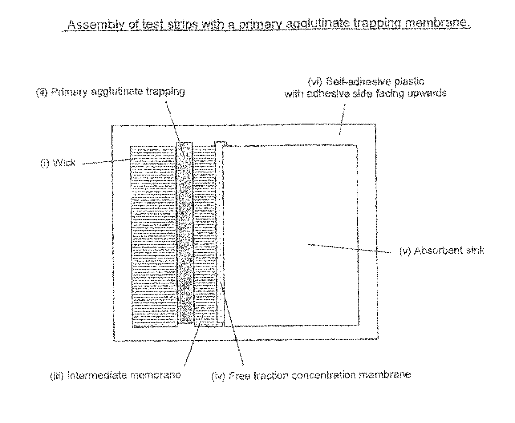
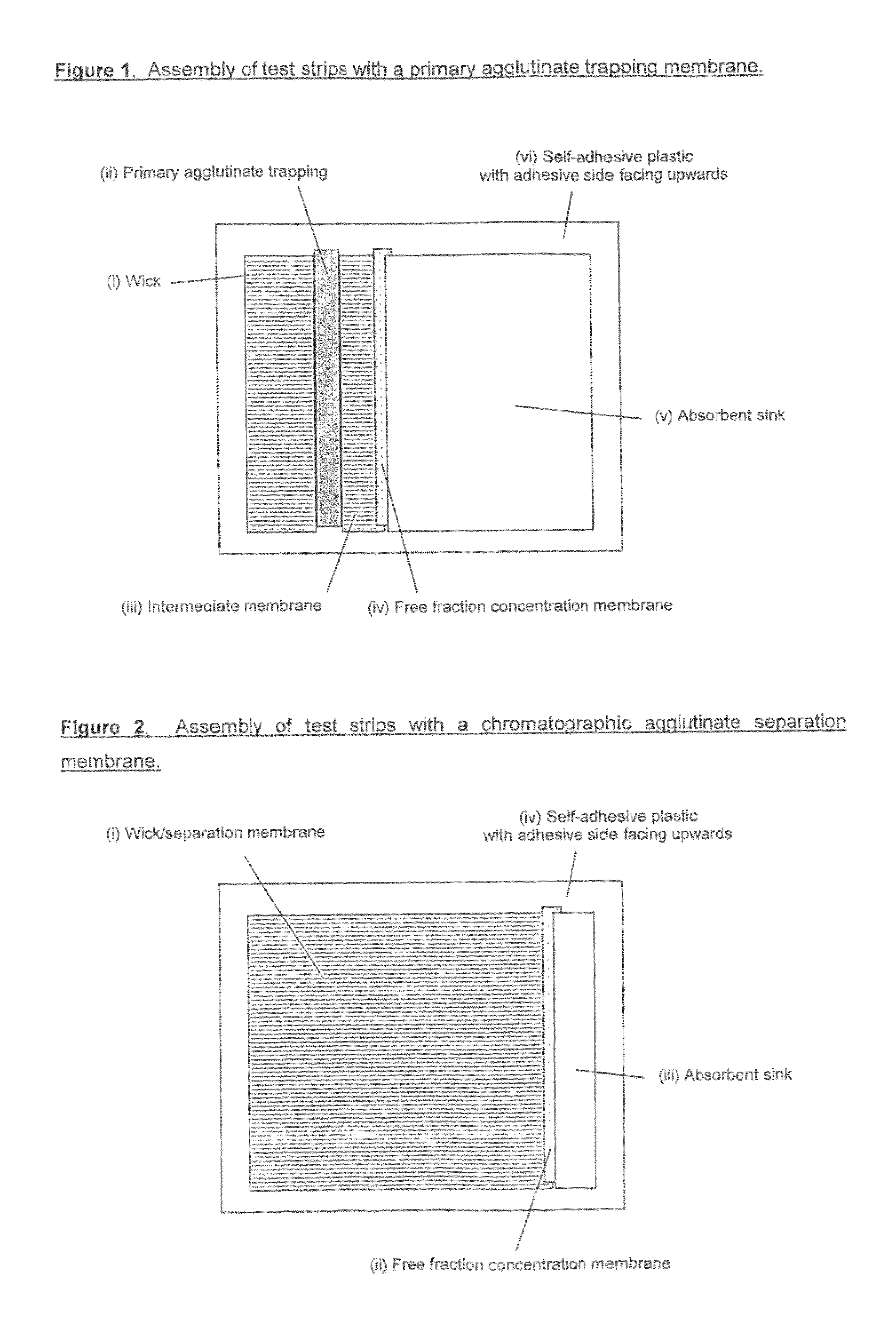
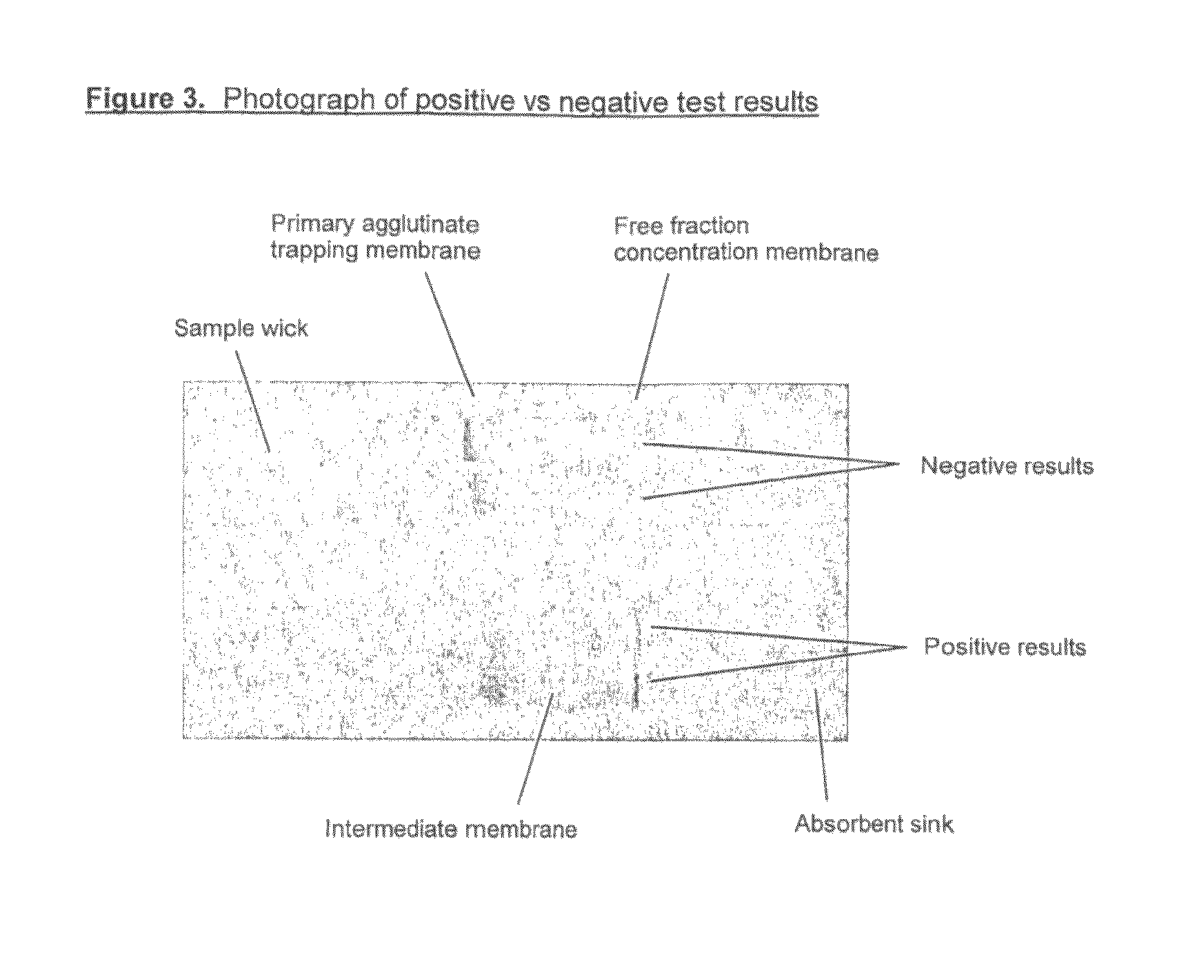
Image

Examples
example 1
Preparation of Antibody-Coated Polystyrene Microparticles
[0097]1. Combine 100 μl 200nm blue polystyrene microparticles 10% solids (‘latex’) (Polymer Labs, Shropshire, UK), 200 μl absolute ethanol, 43 μl sheep FITC antiserum (Micropharm, Carmarthenshire, UK) and 657 μl 10 mM phosphate buffer pH 7.4.[0098]2. Incubate at room temperature for 2 hours, with gentle agitation, to allow antibody adsorption to occur.[0099]3. Add 200 μl 6% BSA in 10 mM phosphate buffer pH 7.4 and continue incubation with agitation for 1 hour.[0100]4. Centrifuge adsorption mixtures for 20 minutes at 4000 g, followed by 5 minutes at 8000 g, to form a soft latex pellet.[0101]5. Remove supernatant and replace with 1 ml latex dilution buffer (Omega Diagnostics, Alva, UK). Gently resuspend pellet.[0102]6. Collect latex pellet by centrifugation and wash as described in 5.[0103]7. Repeat step 6 twice further and finally resuspend latex pellet in 900 μl latex dilution buffer.[0104]8. Adjust latex solids to 1% w / v, by ...
example 2
Preparation of Antibody-Coated Polystyrene Microparticles
[0106]1. Combine 100 μl 200 nm blue polystyrene microparticles 10% solids (‘latex’) (Polymer Labs, Shropshire, UK), 200 μl absolute ethanol, 750 μg mouse IgG (Sigma, 15381) in 10 mM phosphate-buffered saline pH 7.4, 110 μg anti-hCG (Medix Biochemica, Kauniainen, Finland, clone #5006) in 10 mM phosphate-buffered saline pH 7.4. Adjust volume to 1 ml with 10 mM phosphate buffer pH 7.4.[0107]2. Incubate at room temperature for 2 hours, with gentle agitation, to allow antibody adsorption to occur.[0108]3. Add 200 μl 6% BSA in 10 mM phosphate buffer pH 7.4 and continue incubation with agitation for 1 hour.[0109]4. Centrifuge adsorption mixtures for 10 minutes at 3500 g to form a soft latex pellet.[0110]5. Remove supernatant and replace with 1 ml latex dilution buffer (Omega Diagnostics, Alva, UK). Gently resuspend pellet.[0111]6. Collect latex pellet by centrifugation for 20 minutes at 8000 g and wash pellet as described in 5.[0112]...
example 3
Preparation of hCG Hub Reagent
[0115]1. Desalt the anti-hCG (alpha-subunit) into 0.1 M phosphate pH 7.5 buffer, using a 1.6×15 cm G25M Sephadex column, and determine concentration and yield.[0116]2. Activate the anti-hCG antibody, using 8 molar equivalents of NHS-PEG-MAL. Incubate the reaction mixture at 20° C. for two hours. Quench the reaction with 100 molar equivalents of glycine and desalt the maleimide-activated anti-hCG into 5 mM EDTA, PBS pH 7.3 buffer using two shots down a 1.6×15 cm G50F Sephadex column. Determine concentration and yield of activated antibody.[0117]3. Activate a 500 kDa aminodextran using 1000 molar equivalents of 2-Iminothiolane (2-IT). Incubate the reaction mixture at 20° C. for 110 minutes. Desalt the thiol activated aminodextran into 5 mM EDTA, PBS pH 7.3 buffer, using G25M Sephadex media. Determine incorporation ratio of thiol:aminodextran using the Ellman's assay.[0118]4. Add 25 Molar equivalents of the maleimide-activated anti-hCG antibody to the thio...
PUM
| Property | Measurement | Unit |
|---|---|---|
| diameter | aaaaa | aaaaa |
| diameter | aaaaa | aaaaa |
| pH | aaaaa | aaaaa |
Abstract
Description
Claims
Application Information