Detection of bacteria and fungi
a technology of microorganisms and detection methods, applied in the field of detection of microorganisms, can solve the problems of requiring several days to complete the technique, and achieve the effect of facilitating scale up and minimising the amount of sample required
- Summary
- Abstract
- Description
- Claims
- Application Information
AI Technical Summary
Benefits of technology
Problems solved by technology
Method used
Image
Examples
example 2
Demonstration of the Inactivation of Host ATP-Dependent Ligase with NaOH
[0164]Rationale. This experiment was performed in order to demonstrate the ability of alkali pH to inactivate host ATP-dependent ligase released from mammalian white blood cells.
Method
For Mammalian Cells
[0165]45 10 ml of blood was diluted to 50 ml with water to lyse the red cells.
[0166]White cells were collected by centrifugation.
[0167]The cells were resuspended in 50 mM hepes pH 7 and lysed by ribolysis as described in step 15 of example 1 above then diluted 100-fold in water.
[0168]One aliquot of the lysed cells was treated with 5 mM NaOH pH 12 for 20 min whereas another aliquot remained untreated. After treatment with NaOH the lysed cells were diluted into ligase mix and tested for ligase activity as described above in example 1.
For Bacteria
[0169]Cultured E. coli was diluted in water and either treated with 5 mM NaOH, 5 mM NaOH and 50% (v / v) BPer (Fisher Cat. No. 78243) (to lyse the bacterial cells) or with Bp...
example 3
[0178]The purpose of this experiment is to show that yeast (Candida albicans as example) can be detected sensitively even in the presence of blood broth.
[0179]Preparation of Assays Solutions and Components of the Substrate were as Listed Above in Example 1.
Assay Protocol
[0180]A typical assay protocol is as follows.[0181]1. To 0.25 ml 10% (v / v) Triton X-100 in a 15 ml centrifuge tube, add 10 ml blood:broth and mix. Note: If spiking with bacteria or fungi, add them at this step.[0182]2. Incubate for 5 min on the bench then centrifuge 3-4000×g for 20 min.[0183]3. Pour off the supernatant and invert tube on a tissue to dry.[0184]4. Add 1 ml H2O and pipette to resuspend.[0185]5. Add 9 ml H2O and invert to mix. Add 1 ml 50 mM NaOH and invert to mix[0186]6. Incubate 5 min on the bench then centrifuge 3-4000×g for 20 min.[0187]7. Pour off supernatant and invert tube to dry.[0188]8. Resuspend pellet in 1 ml 50 mM Tris pH 7.5, transfer to microfuge tube, spin 8,000 ...
experiment 1
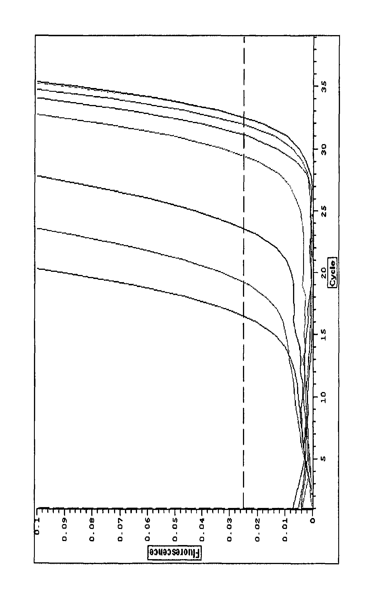
[0198]C. albicans in culture medium vs C. albicans in blood broth (NaOH treated).
[0199]When C. albicans was measured using the above protocol, with an NaOH treatment step, the results were as shown in Table 1 below:
[0200]
TABLE 1Culture mediumBlood brothNumericalNumericaldifferencedifferenceCtfromCtfromNumber ofdiffer-controldiffer-controlC. albicansCtence(fold)Ctence(fold)390 CFU / mL 24.13.511.324.54.624.398 CFU / mL26.11.52.827.02.14.325 CFU / mL25.32.34.927.71.42.6Control27.6029.10
[0201]Because each Ct difference represents a two-fold increase in the signal, the figures in the “numerical difference” column are given to show the actual difference. For example, 390 CFU / mL C albicans gave an 11.3-fold increase in signal over background or a 3.5Ct difference in culture medium.
[0202]The results show:[0203]1. C. albicans can be measured sensitively in blood broth.[0204]2. The background signal in blood broth is very low when the NaOH treatment has been used.
PUM
| Property | Measurement | Unit |
|---|---|---|
| pH | aaaaa | aaaaa |
| pH | aaaaa | aaaaa |
| temperatures | aaaaa | aaaaa |
Abstract
Description
Claims
Application Information
 - R&D
- Intellectual Property
- Life Sciences
- Materials
- Tech Scout
- Unparalleled Data Quality
- Higher Quality Content
- 60% Fewer Hallucinations
Browse by: Latest US Patents, China's latest patents, Technical Efficacy Thesaurus, Application Domain, Technology Topic, Popular Technical Reports.
© 2025 PatSnap. All rights reserved.Legal|Privacy policy|Modern Slavery Act Transparency Statement|Sitemap|About US| Contact US: help@patsnap.com

