Multi energy X-ray microscope data acquisition and image reconstruction system and method

a multi-energy x-ray microscope and data acquisition technology, applied in the field of multi-energy x-ray microscope data acquisition and image reconstruction system and method, can solve the problem of no convenient way to combine and analyze data, and achieve the effect of improving contrast and image quality

Active Publication Date: 2015-09-08
CARL ZEISS X RAY MICROSCOPY
View PDF6 Cites 11 Cited by
  • Summary
  • Abstract
  • Description
  • Claims
  • Application Information

AI Technical Summary

Benefits of technology

[0014]The present invention provides a multi energy, such as dual-energy, x-ray imaging system data acquisition and image reconstruction system and method for combining separate reconstructed volumes taken at multiple energies and allowing the operator to manipulate and optimize the image contrast of a sample using the effects of DE. Operators use the system and method to achieve superior image quality and contrast over current XRM data acquisition and image reconstruction systems and methods, and to generate contrast based on the atomic number Z of the constituents of the sample.
[0016]The system subsequently performs a registration, zooming, and scaling step of the two separately reconstructed tomographic volumes to adjust for imperfections in the spatial stretch and relative position of the low-energy and high-energy tomographic datasets. This ensures the correlation of voxels in both datasets.
[0018]The two-dimensional histogram tool plots the intensity of pixels of the low energy slice against the intensity of pixels of the high energy slice. The intensity in the two dimensional histogram represents the number of voxels that fall into the corresponding histogram bin. The intensity is visualized by user-selectable color-maps that aid in the visibility and interpretation of the histogram.
[0021]In addition to optimizing image contrast for features within a sample, usage of dual-energy can result in faster total acquisition and reconstruction time than that obtained by standard absorption techniques for certain samples.
[0027]It is another aspect to provide optimum single scan parameters from information associated with the DE contrast-optimized volume data set to best approximate the operator-selected settings for the DE contrast-optimized volume data set. In this way, an operator can run subsequent single-energy scans that provide improved contrast and image quality as compared to volume data sets created from current single-energy scanning methods.

Problems solved by technology

One limitation of current XRMs and their associated imaging methods is that there is no convenient way to combine and analyze the data after creating the reconstructed volume data sets for the energies for the sample.

Method used


Image

  • Multi energy X-ray microscope data acquisition and image reconstruction system and method
  • Multi energy X-ray microscope data acquisition and image reconstruction system and method
  • Multi energy X-ray microscope data acquisition and image reconstruction system and method

Examples


Embodiment Construction

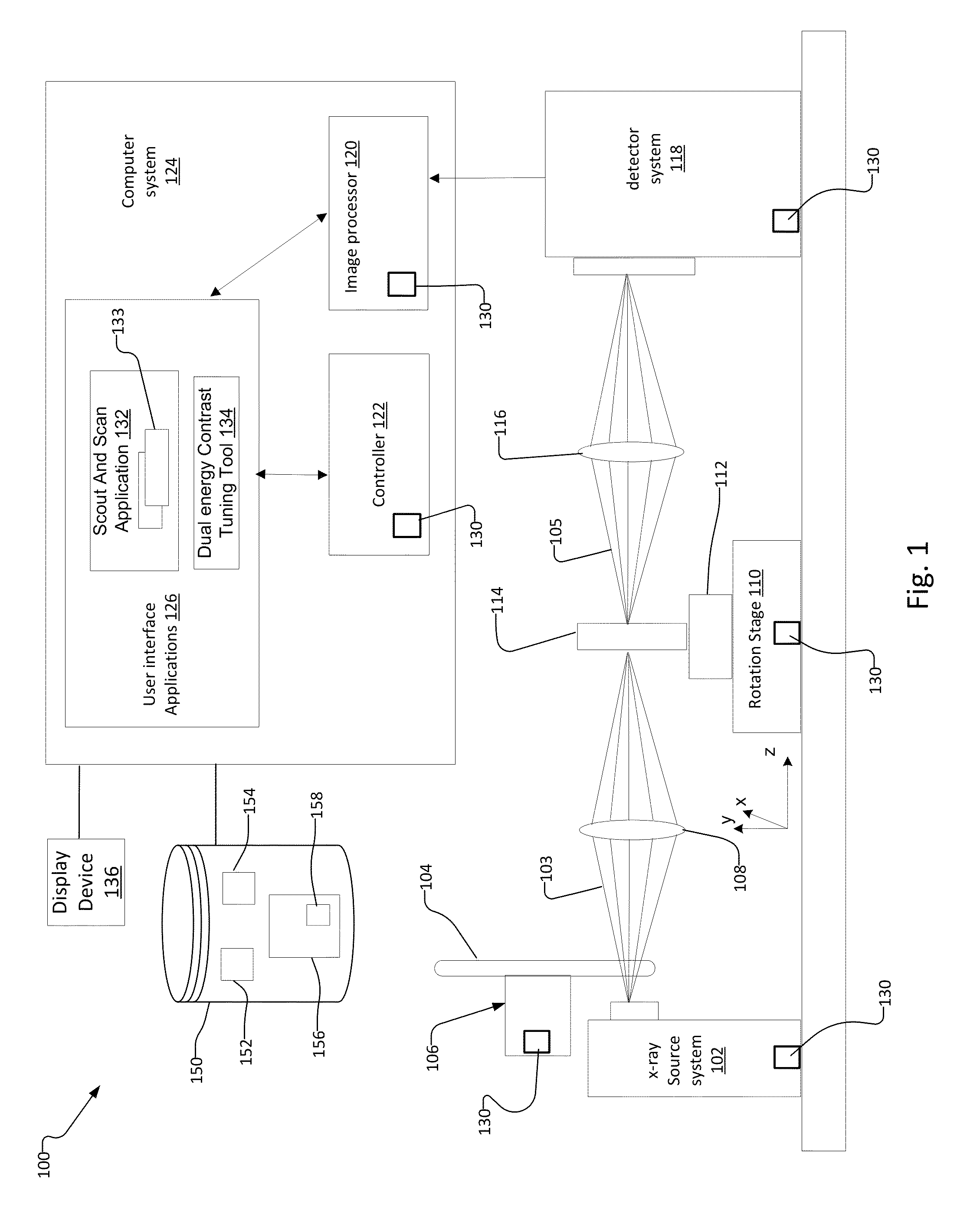
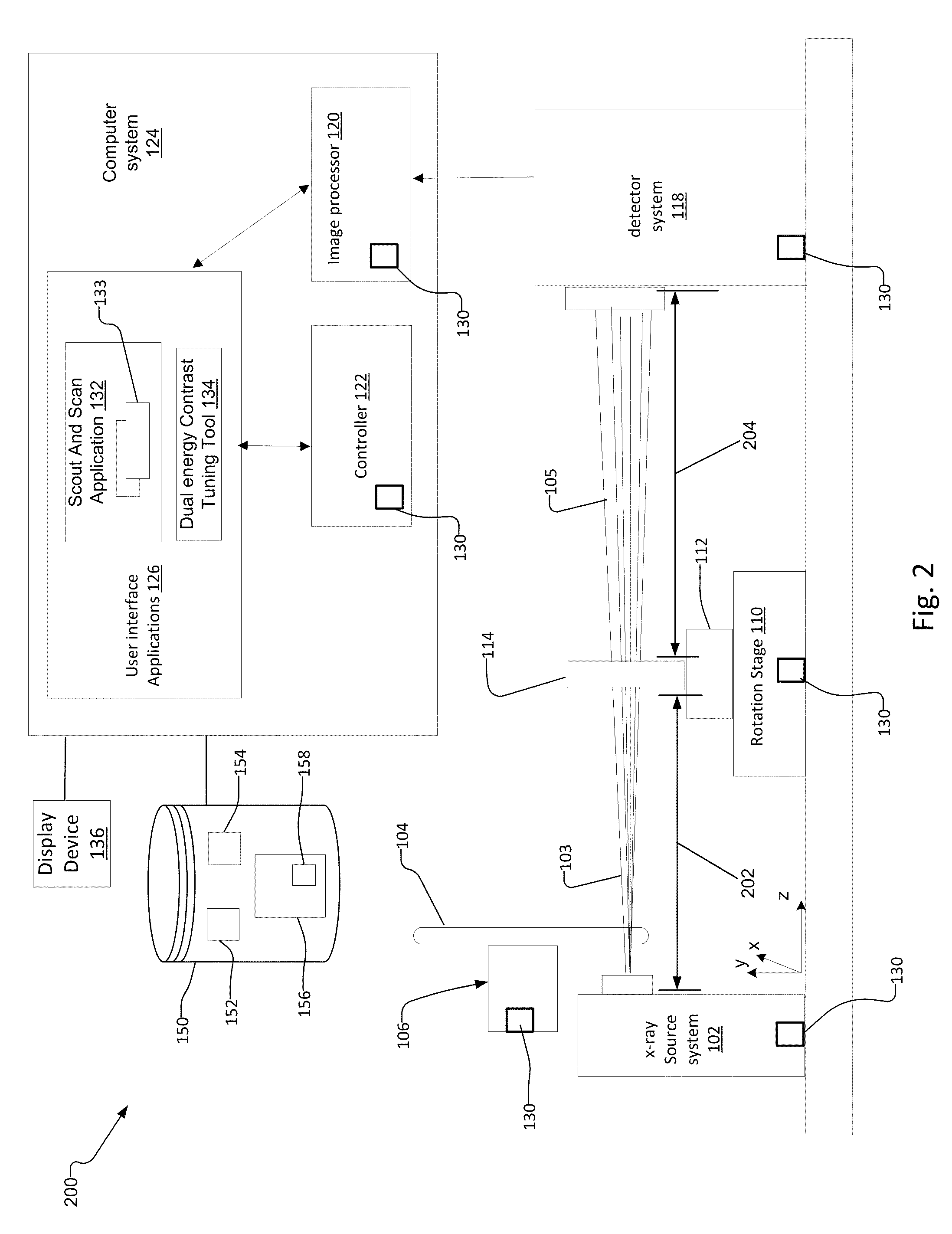
[0046]FIG. 1 is a schematic diagram of a lens-based x-ray imaging system 100 (“lens-based system”).

[0047]The lens-based system 100 has an x-ray source system 102 that generates an x-ray beam 103, a filter changer mechanism 106 with a filter wheel 104 for filtering the x-ray beam 103, and a rotation stage 110 with sample holder 112 for holding the sample 114. A condenser 108 placed between the x-ray source system 102 and the sample 114 focuses the x-ray beam 103 onto the sample 114.

[0048]The lens-based system 100 also has a detector system 118, and an objective lens 116 placed between the sample 114 and the detector system 118. When the sample 114 is exposed to the x-ray beam 103, the sample 114 absorbs and transmits x-ray photons associated with the x-ray beam 103. The x-ray photons transmitted through the sample form an attenuated x-ray beam 105, which the objective lens 116 images onto the detector system 118.

[0049]The detector system 118 creates an image representation, in pixels...


PUM

PropertyMeasurementUnit
energyaaaaaaaaaa
volumeaaaaaaaaaa
tomographic volumeaaaaaaaaaa

Abstract

A multi energy, such as dual-energy (“DE”), x-ray imaging system data acquisition and image reconstruction system and method enables optimizing the image contrast of a sample. Using the DE x-ray imaging system and its associated user interface applications, an operator performs a low energy (“LE”) and high energy (“HE”) x-ray scan of the same volume of interest of the sample. The system creates a low-energy reconstructed tomographic volume data set from the set of low-energy projections and a high-energy tomographic volume data set from the set of high-energy projections. This enables the operator to control the image contrast of selected slices, and apply the information associated with optimizing the contrast of the selected slice to all slices in the low-energy and high-energy tomographic data sets. This creates a combined volume data set from the LE and HE volume data sets with optimized image contrast throughout.

Description

[0001]A portion of the disclosure of this patent document contains material that is subject to copyright protection. The copyright owner has no objection to the facsimile reproduction by anyone of the patent document or the patent disclosure, as it appears in the Patent and Trademark Office patent file or records, but otherwise reserves all copyright rights whatsoever.BACKGROUND OF THE INVENTION[0002]High resolution x-ray imaging systems, also known as X-ray imaging microscopes (“XRM”), provide high-resolution / high magnification, non-destructive imaging of internal structures in samples for a variety of industrial and research applications, such as materials science, clinical research, and failure analysis to list a few examples. XRMs provide the ability to visualize features in samples without the need to cut and slice the samples. XRMs are part of the field of x-ray microscopy.[0003]XRMs are often used to perform computed tomography (“CT”) scans of samples. CT scanning is the proc...

Claims


Application Information

Patent Timeline
Patent Type & Authority Patents(United States)
IPC IPC(8): G01N23/087G21K7/00G06T11/00G06F3/0481A61B6/00G01N23/04G06K9/62A61B6/03G06T5/40G06T5/50G06T15/08
CPCG06F3/0481A61B6/482G01N23/046G06K9/6212G06T5/40G06T5/50G06T11/006G21K7/00A61B6/03A61B6/52G01N2223/419G01N2223/423G06T15/08G06F3/04842
Inventor CASE, THOMAS A.CANDELL, SUSANSESHADRI, SRIVATSANMCGUINNESS, PAUL
Owner CARL ZEISS X RAY MICROSCOPY
Why Eureka
Try Eureka