Quantitative assessment of neovascularization

a neovascularization and quantitative technology, applied in the field of quantitative assessment of neovascularization in plaques, can solve the problems of spatial resolution, imaging tools such as ct, mri and pet-spect, cannot reasonably resolve such small diameters, etc., and achieve more reproducible results, improve accuracy, and enhance probability

Inactive Publication Date: 2015-12-22
TECHNION RES & DEV FOUND LTD
View PDF15 Cites 3 Cited by
  • Summary
  • Abstract
  • Description
  • Claims
  • Application Information

AI Technical Summary

Benefits of technology

[0011]The contrast enhanced ultrasound technique involves the injection of a contrast enhancing medium into the patient's blood stream. The effect of the impinging ultrasound on such a contrast medium is to activate the small gas bubbles, typically of a 1-3 μm in diameter, and these reflect the ultrasound waves efficiently, whereas the blood itself does not. However, the contrast enhancing agent is spread out into discrete and isolated locations in the blood stream, such that the blood vessel in which it is imaged appears like an intermittently dashed line. The main reason for the discrete blobs may be the very high resistance of the tiny intra-plaque neovessels. The system and methods described in this disclosure are capable of distinguishing between the ultrasound reflections from the contrast material flowing in these small blood vessels, and the ultrasound reflections from the surrounding tissue due to harmonic imaging. The body tissues generate echoes at mostly the fundamental frequency of the transmitted ultrasound frequency (in harmony with the original transmitted frequency). On the other hand, the contrast agents, due to their nonlinear behavior, can also produce sub-harmonic and second-harmonic frequencies, thus enabling imaging of the contrast enhancing medium and reconstruction of its flow path.
[0017]Several logical decision procedures may be used to enhance the probability that several segments or pixels / volumes are positively included within a branch of the arteriole tree. The area and / or volume of blood / contrast agent contained within the lumen of the arterioles that form the arteriole tree is then estimated. In addition, the orientation of the flow over time is calculated, as part of the delineation process of the shape of the arteriole tree. As a result of executing the above described steps, a description may be obtained, in 2D or 3D, of the arteriole tree within the ROI of the tumor or plaque, together with the calculated volume of that tree. The reconstruction of a 3D arterial tree may be more accurate than the 2D analysis because it is less dependent in the imaged cross-section and in the location of the ultrasound probe, thus supplying more reproducible results. The process includes monitoring of different 2D cross-sections of the plaque or tumor, and reconstructing those sections over a whole area. Additionally, it is possible to obtain shape parameters such as the length of the branches and the branching morphology of the vessels within the tumor or a plaque.
[0018]The above described method, in all of its implementations, can be utilized as part of the image processing routines of the ultrasound imaging system, or as a stand-alone post-processing package, and provides clinicians with novel screening and monitoring capabilities in the diagnosis and treatment of disease. It allows clinicians who diagnose or treat patients with cancerous tumors to assess the potential rate of growth of the tumor, and its response to anti-angionetic therapy; it allows clinicians who assess the vulnerability of plaques to do so in a quantitative manner; and it allows for the screening of patients who may be susceptible to stroke or acute myocardial infarction.

Problems solved by technology

In the first place, a problem of spatial resolution exists.
The common imaging tools used, such as CT, MRI and Pet-Spect, can not reasonably resolve such small diameters.

Method used


Image

  • Quantitative assessment of neovascularization
  • Quantitative assessment of neovascularization
  • Quantitative assessment of neovascularization

Examples


Embodiment Construction

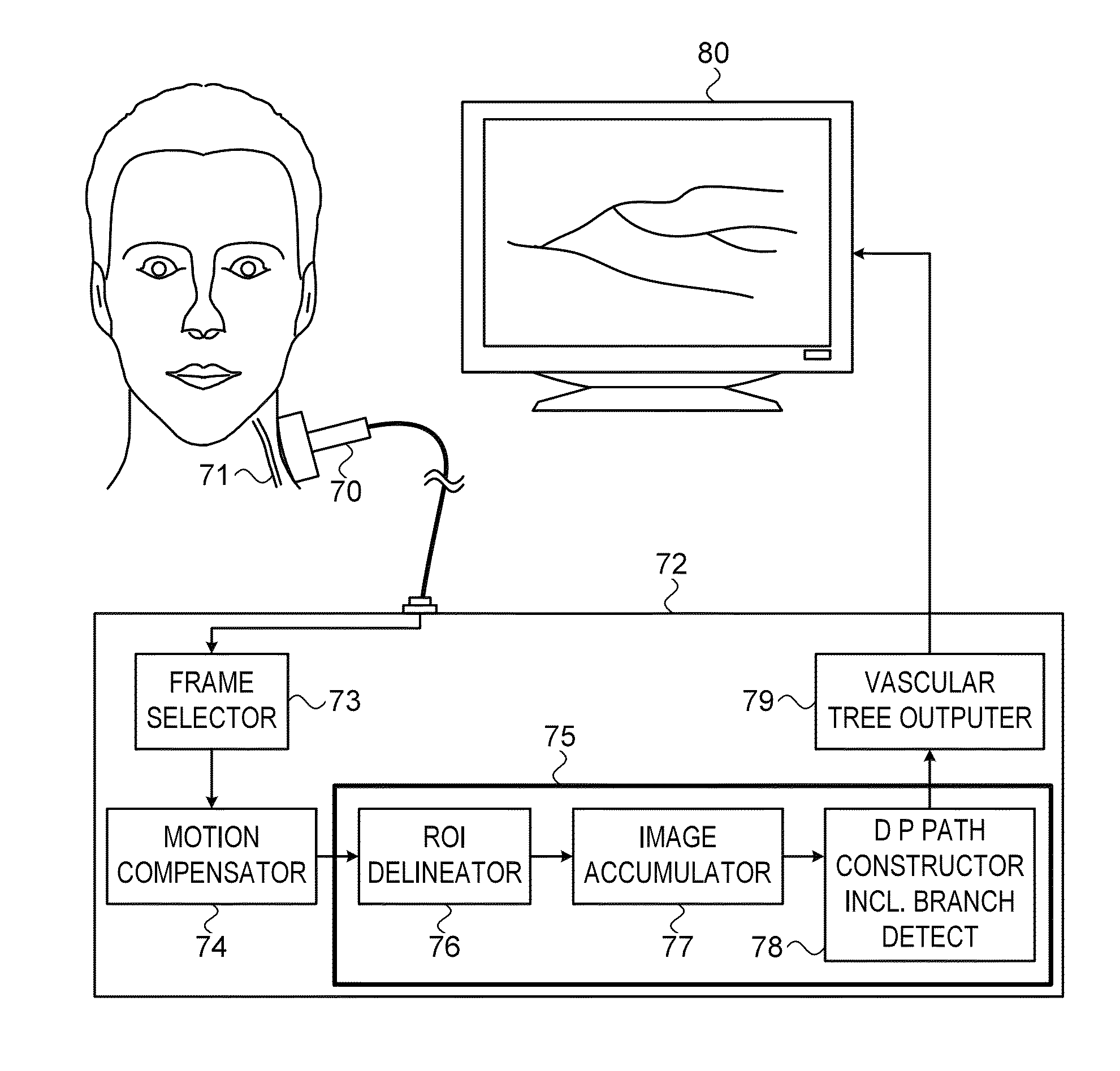
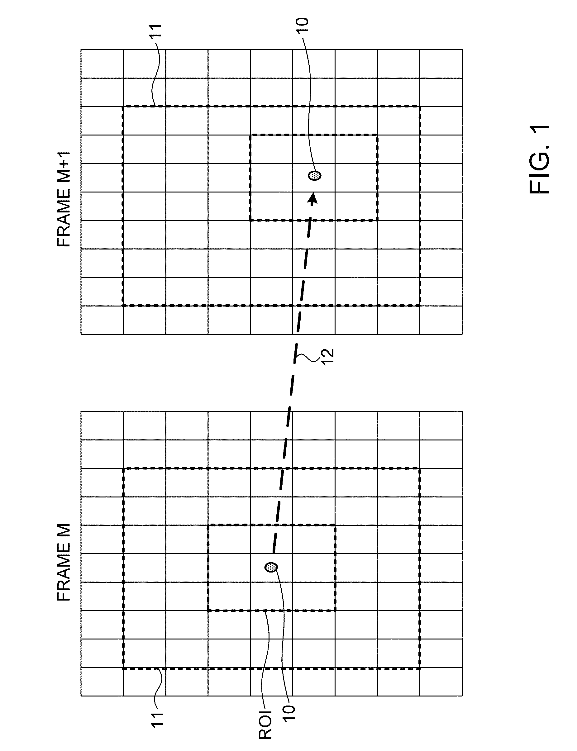
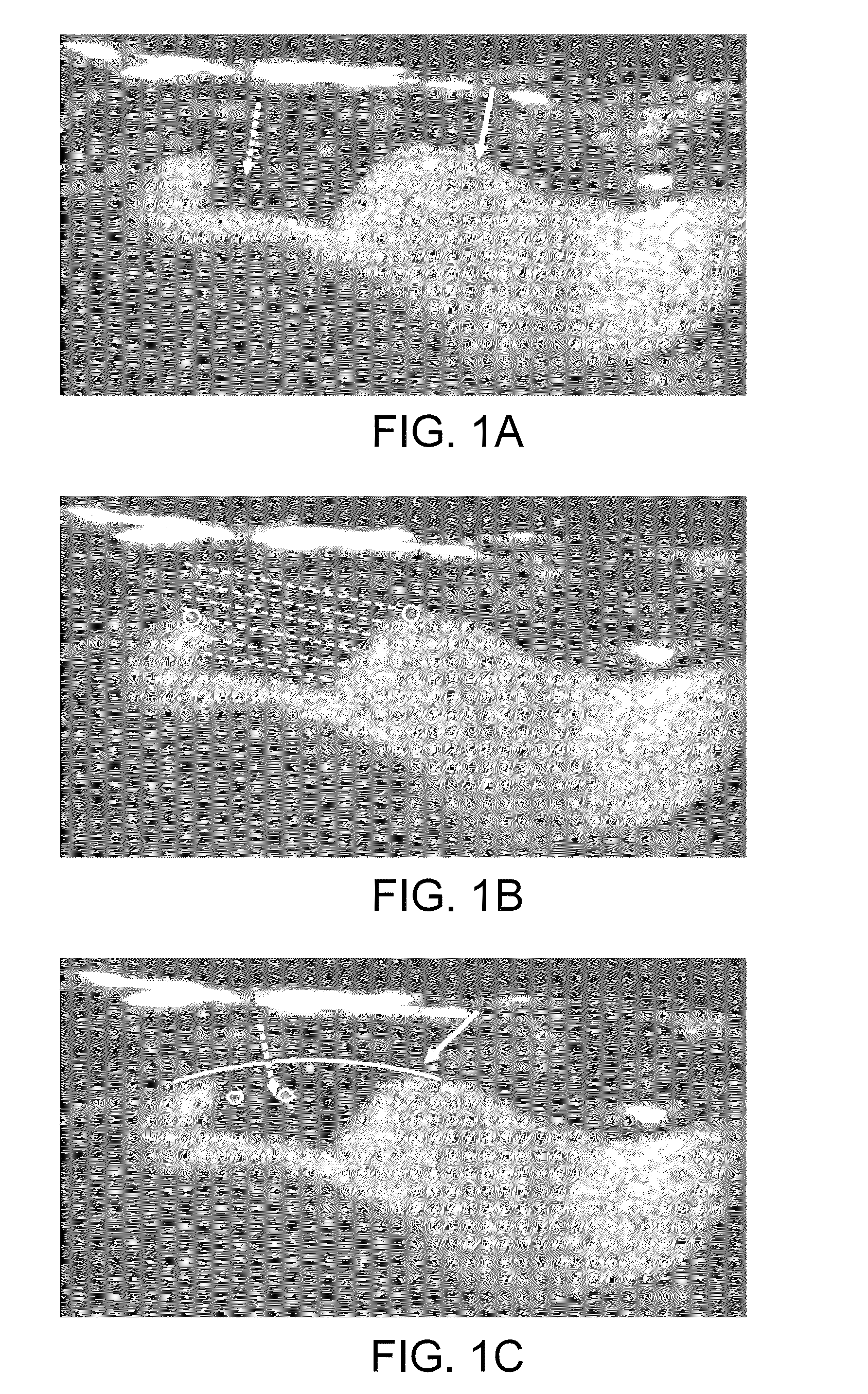
[0040]Exemplary systems and methods for quantification of neovascularization, as described in the present disclosure, involve the acquisition of contrast enhanced ultrasound (CEUS) images in the region where the neovascularization is to be calculated. Ultrasound examination of the carotid bifurcation and internal carotid artery is used as an exemplary application of the methods of this disclosure. Typically, a routine ultrasound grayscale image procedure is performed to assess the position, size and shape of the plaque (if neovascularization in a plaque is being measured), followed by color and spectral Doppler imaging to confirm the degree of stenosis. This can be performed, for instance, on an iU22 ultrasound system, available from Philips Healthcare of Bothwell, Wash., with a 4-8 MHz broad band linear array transducer. Thereafter the activated contrast agent, such as Definity™ as supplied by Bristol-Myers Squibb Medical Imaging Corp., of North Billerica, Mass., diluted in saline,...


PUM


Abstract

Systems and methods using contrast enhanced ultrasound imaging for quantitative assessment of neovascularization, such as within a tumor or a plaque. The contrast agent moves in small blotches rather than in continuous flow, but image processing methods enable the detection and quantification of the blood flow, even in the tiny blood vessels of the vasculature tree. The image processing includes compensation for motion of the tissue arising from body movement. The position and extent of the discrete positions of contrast material may be accumulating during a complete heart cycle onto a single 2D image, to enable an indication of a vascular tree based on this local accumulated image. Dynamic processing (DP) of the discrete positions of contrast material enables a complete vascular tree to be obtained. The neovascularization is quantified by the ratio of its total area to that of the plaque in which the neovascularization has grown.

Description

CROSS-REFERENCE TO RELATED APPLICATIONS[0001]This application claims the benefit of priority from U.S. Provisional Patent Application No. 61 / 592,068, filed Jan. 30, 2012, the contents of which is incorporated herein by reference.FIELD OF THE INVENTION[0002]The present invention relates to the field of the quantification of neovascularization in plaque or in tumors, especially using contrast enhanced ultrasound.BACKGROUND OF THE INVENTION[0003]Neovascularization is the formation of new blood vessels—generally capillary ingrowth or endothelial proliferation—in unusual sites. The phenomenon is typical of so-called angiogenic diseases, which include angiogenesis in tumor growth. Recently, it has been determined that neovascularization can also occur in arterial plaques. Thus, during atherosclerosis development, reduction of the oxygen supply to the vessel wall may occur, resulting in ischemia, which induces a continual release of growth factors that stimulate the neovascularization proc...

Claims


Application Information

Patent Timeline
Patent Type & Authority Patents(United States)
IPC IPC(8): A61B8/00A61B8/08G06T7/00A61B8/06
CPCA61B8/0891A61B8/085A61B8/481A61B8/5207A61B8/5276G06T7/0012G06T2207/30101A61B8/06G06T2207/10132G06T2207/20221G06T2207/30096
Inventor HOOGI, ASSAFBAR-ZION, AVINOAMADAM, DAN
Owner TECHNION RES & DEV FOUND LTD
Why Eureka
Try Eureka