Brachytherapy dose verification apparatus, system and method

a brachytherapy and dose technology, applied in the field of system, method and device for brachytherapy treatment verification, can solve the problems of insufficient brachytherapy dose verification, inability to accurately determine the source calibration data of the brachytherapy treatment console, complex hdr brachytherapy treatment, etc., and achieve the effect of increasing the image array

Inactive Publication Date: 2017-05-02
SMITH RYAN LEE +1
View PDF30 Cites 1 Cited by
  • Summary
  • Abstract
  • Description
  • Claims
  • Application Information

AI Technical Summary

Benefits of technology

[0256]According to the sixth aspect the distance between the brachytherapy radiation...

Problems solved by technology

HDR brachytherapy treatment can be complex, usually requiring computed tomography (CT) data to accurately plan the treatment on a specialised treatment planning system (TPS).
Inaccuracies in treatment delivery can occur due to several reasons; implanted catheters move inside the patient after CT imaging1, specified length of the catheter is incorrect, catheter tip offset is incorrect, transfer tube connection is incorrect (afterloader to patient catheter), wrong treatment plan is delivered, remote afterloader malfunctions, source calibration data in the brachytherapy treatment console is incorrect2.
A limitation of this method...

Method used


Image

  • Brachytherapy dose verification apparatus, system and method
  • Brachytherapy dose verification apparatus, system and method
  • Brachytherapy dose verification apparatus, system and method

Examples


examples

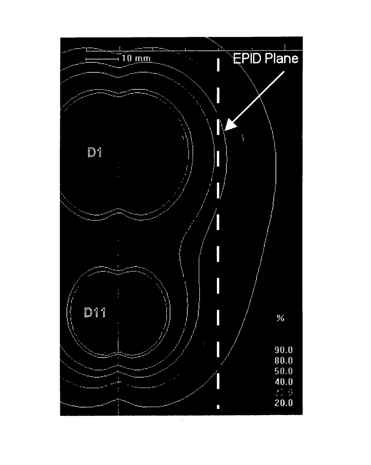
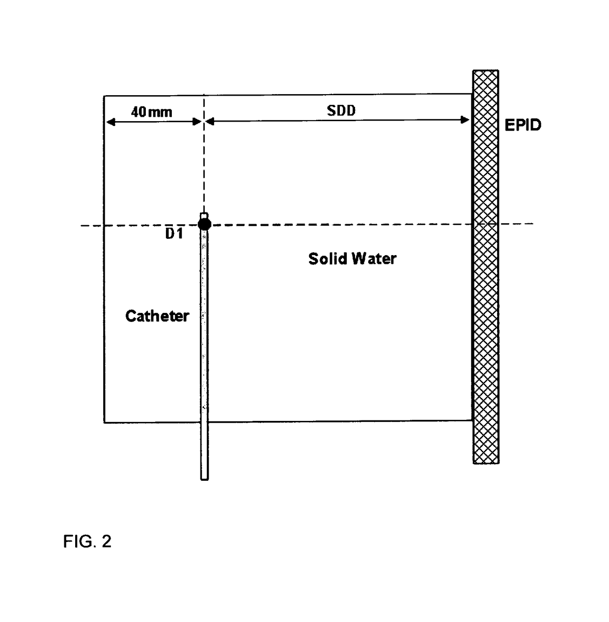
[0537]The EPID used throughout all experiments was an amorphous silicon flat panel imager (Varian Medical Systems, Palo Alto, Calif.), detector model IAS11-19 running software version 6.1.11. Normally fitted to an external beam linear accelerator, the detector was removed and remained mobile for all measurements. The imager is operated using the AM Maintenance software module (version 7.1.2003.905). The EPID has a 400×300 mm2 detection area (512×384 pixels), a 1 mm Cu build-up layer, a phosphor screen and a hydrogenated a-Si:H photodiode array. The touch guard cover was removed for all measurements. Further details regarding similar types of imagers can be found in an overview by Antonuk et al.18

[0538]The stored EPID image is the average of all frames captured between the start and stop signals that define an image acquisition. Each frame is defined as the signal from one readout of the entire photodiode array. This average of all frames, makes the interpretation of EPID output, as ...


PUM


Abstract

A system, method and device for brachytherapy treatment verification is described herein. The verification may be in real time and may provide verification of one or more of dose, source position, dwell time and source activity. In one embodiment the invention provides a method for verifying a brachytherapy radiation treatment including processing a distribution of exposure to a brachytherapy radiation source of a two dimensional imaging array to determine a region of high exposure; obtaining one or more distribution of exposure profiles through the region of high exposure; determining a region of high value in the one or more distribution of exposure profiles; and using the determined region of high exposure and/or high value to calculate one or more brachytherapy radiation source position and/or one or more brachytherapy radiation source distance to thereby verify at least a part of the brachytherapy radiation treatment.

Description

CROSS REFERENCE TO RELATED APPLICATIONS[0001]This is the U.S. National Stage of International Application No. PCT / AU2010 / 001188, filed Sep. 13, 2010, which was published in English under PCT Article 21(2).FIELD[0002]THIS INVENTION described herein relates generally to a system, method and device for brachytherapy treatment verification. In particular, the invention is directed to, a system, method and device for real time brachytherapy treatment verification. The treatment verification may be in real time and may provide verification of one or more of dose, source position, dwell time and source activity although the scope of the invention is not necessarily limited thereto.BACKGROUND[0003]High Dose Rate (HDR) brachytherapy is a form of radiotherapy for cancer treatment that can deliver a high dose of radiation to the target volume, while minimising dose to the nearby healthy tissue. HDR brachytherapy is delivered by one or more high activity sources, moved through catheters placed ...

Claims


Application Information

Patent Timeline
IPC IPC(8): A61N5/10A61B6/12
CPCA61N5/1071A61B6/12A61N5/1001A61N5/1064A61N2005/1054A61N2005/1072
Inventor SMITH, RYAN LEEFRANICH, RICK DON
Owner SMITH RYAN LEE
Why Eureka
Try Eureka