Device combining magnetic resonance imaging and positron emission tomography for breast examination

a technology of magnetic resonance imaging and positron emission tomography, which is applied in the direction of patient positioning for diagnostics, measurement using nmr, instruments, etc., can solve the problems of high cost of two imaging examinations, inability to show anatomy and detailed, and need for further correction of images, so as to reduce the friction of the first nylon rope and the loading weight, increase the actuation speed of the device, and reduce the loading weight and friction

Inactive Publication Date: 2017-05-09
SHIH MING CHANG +1
View PDF5 Cites 0 Cited by
  • Summary
  • Abstract
  • Description
  • Claims
  • Application Information

AI Technical Summary

Benefits of technology

[0008]In the foregoing description, the counterweight unit decreases friction of the first nylon rope and the loading weight of the PET scanner ring by counterweight to increase an actuation speed of the device. The displacement measurement unit is further provided with an optical scale connecting to the second nylon rope and a decoder card for receiving a pulse output from the optical scale and converting it into a location signal.
[0009]In use of a device of the present invention, a subject undergoing breast MRI is made to lie face down on a breast MRI bed with two holes for the breasts to fit in. These holes have a network of magnetic lines for the MRI to take place. Scanning position of the PET bearing platform can be modified by the servo flow control module and the displacement measurement unit. Moreover, the decreased loading weight and friction by the counterweight unit can enhance the speed of operation of the bearing platform. Accordingly, a device combines PET to the breast MRI and takes advantage of a servo flow control module, a non-metallic and non-magnetic pneumatic actuator, a movable pulley and the like to control a position of the bearing platform, so that subjects' breasts can be accurately detected within a short time.

Problems solved by technology

However, there are some disadvantages respectively existed in these two medical imaging techniques, i.e. MRI is unable to offer metabolic information and disease status and PET is in lack of showing anatomy and detailed location.
Moreover, these two imaging examinations cost high and take a lot of subject's time and energy in each inspection.
However, the move of the subject results in the image distortion, so the images need further corrections.
Although Siemens Co. has developed the PET / MRI system for examining subjects' whole bodies, combining a PET with a breast MRI still has a problem, i.e. difficulty in accommodating a PET machine due to the small space of a breast MRI system.

Method used


Image

  • Device combining magnetic resonance imaging and positron emission tomography for breast examination
  • Device combining magnetic resonance imaging and positron emission tomography for breast examination
  • Device combining magnetic resonance imaging and positron emission tomography for breast examination

Examples


Embodiment Construction

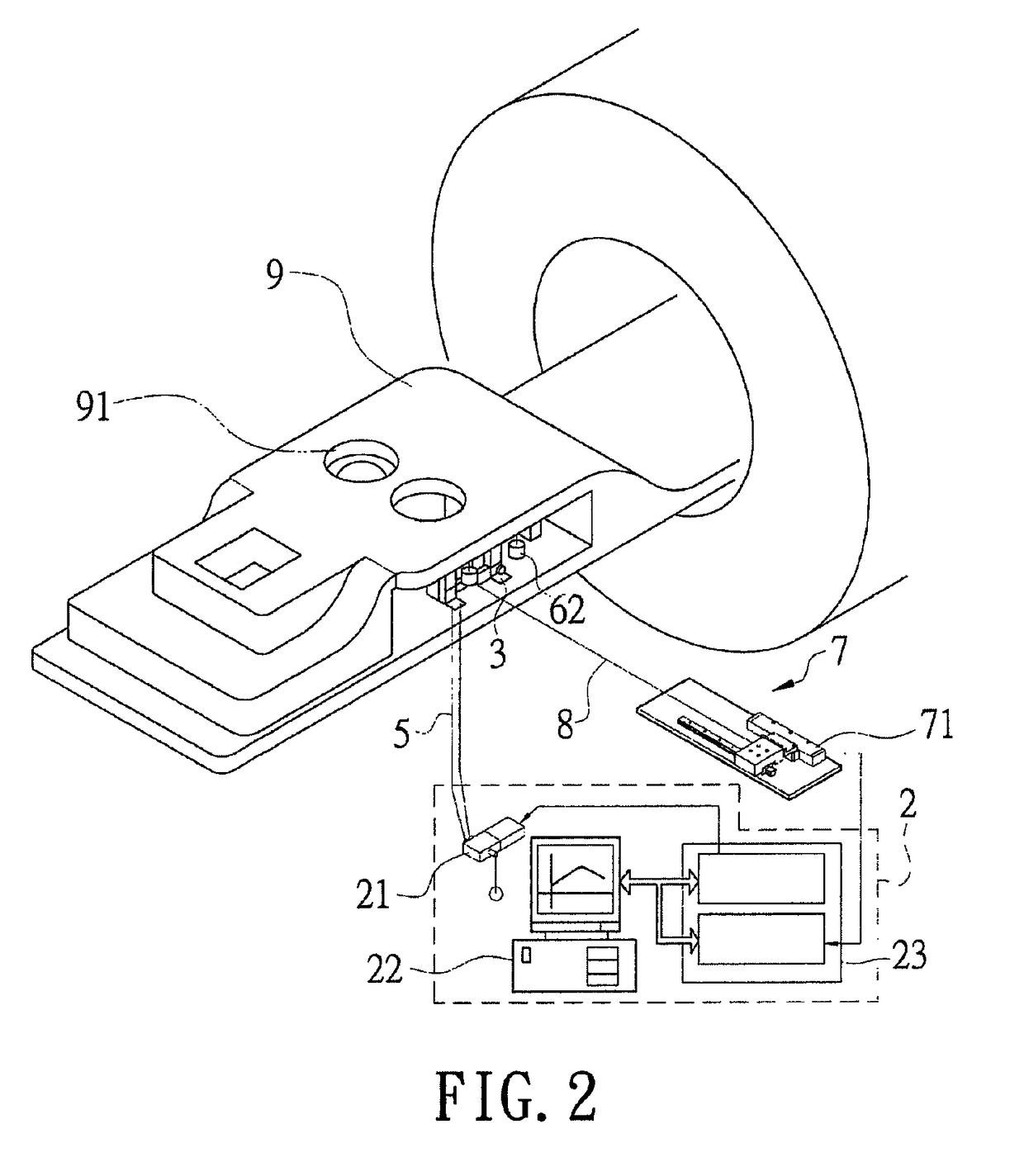
[0014]The present invention relates to a device combining magnetic resonance imaging and positron emission tomography for a breast examination. The device comprises a PET scanner ring disposed in the narrow place of a breast MRI bed by a mechanism design approach, so as to precisely examine the breasts and acquire better images within a short time.

[0015]Hereinafter, an exemplary embodiment of the present invention will be described in detail with reference to the accompanying drawings.

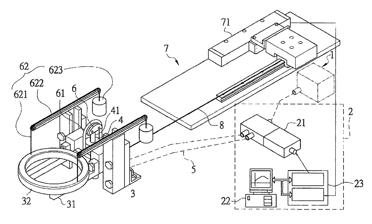
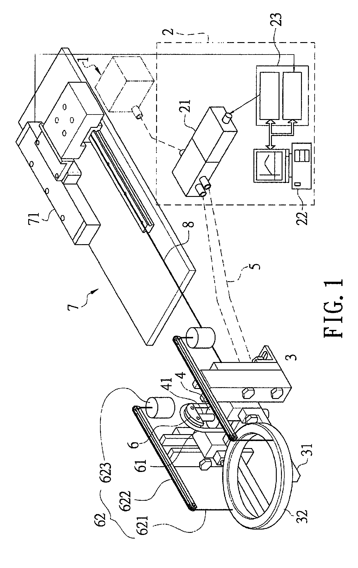
[0016]Referring to FIG. 1, FIG. 2 and FIG. 4, a partially enlarged diagram showing a local device structure according to the present invention, a schematic diagram showing a device combining magnetic resonance imaging and positron emission tomography for a breast examination according to the present invention, and a system diagram showing a device equipped with a counterweight unit according to the present invention are revealed, comprising:

[0017]an air pressure source (1);

[0018]a servo flow control mo...


PUM


Abstract

A device combining magnetic resonance imaging and positron emission tomography for a breast examination is revealed herein to comprise an air pressure source, a servo flow control module, a pedestal disposed under a breast accommodating hole of a breast MRI bed and having a bearing platform for bearing a PET scanner ring thereon, a non-metallic and non-magnetic pneumatic actuator disposed on the pedestal and connected with the servo flow control module, and a movable pulley having a first nylon rope whose one end connects to the bearing platform and the other end connects to a counterweight unit, wherein the bearing platform connects to a displacement measurement unit by a second nylon rope for receipt of a location information thereof and transmission of the same to the servo flow control module for controlling the opening of the servo flow control valve to change gas flows entering into the pneumatic actuator.

Description

BACKGROUND OF THE INVENTION[0001]1. Field of the Invention[0002]The present invention relates to a device combining magnetic resonance imaging (MRI) and positron emission tomography (PET) for a breast examination. The device comprises a PET scanner ring disposed in a narrow place of a breast MRI bed in order to precisely examine the breasts and acquire better images within a short time.[0003]2. Description of Related Art[0004]Generally, both magnetic resonance imaging (MRI) and positron emission tomography (PET) examinations are required for a subject to assist physicians in the clinical diagnosis and judgment. Magnetic resonance imaging is used to investigate the anatomy and detailed location of subjects' bodies, and positron emission tomography (PET) provides information of cell physiology and metabolism. However, there are some disadvantages respectively existed in these two medical imaging techniques, i.e. MRI is unable to offer metabolic information and disease status and PET i...

Claims


Application Information

Patent Timeline
Patent Type & Authority Patents(United States)
IPC IPC(8): G01R33/48A61B6/04A61B6/03A61B6/00
CPCG01R33/481A61B6/037A61B6/0435A61B6/502A61B6/5247
Inventor SHIH, MING-CHANGHUANG, HUNG-LUNG
Owner SHIH MING CHANG
Why Eureka
Try Eureka