Method and system for detecting and/or classifying cancerous cells in a cell sample

a cell sample and cancerous cell technology, applied in the field of diagnostics, can solve the problems of lack of sensitivity, high specificity of pap smear test, and lack of sensitivity, and achieve the effects of non-destructive, fast, inexpensive and objective, and high specificity and sensitiv

Active Publication Date: 2017-06-20
OVIZIO IMAGING SYST NV SA
View PDF12 Cites 20 Cited by
  • Summary
  • Abstract
  • Description
  • Claims
  • Application Information

AI Technical Summary

Benefits of technology

[0010]The present invention provides for a method and system for analyzing cell samples in a non-destructive, fast, inexpensive and objective manner and to detect cancerous cells present in the sample. In the current invention, said cell sample will be analyzed by a digital holographic microscope (DHM) and the practitioner will be provided with a digital report, comprising a set of cellular parameters related to cells present in the cell sample as well as with diagnostic information on the cell sample. This will give the practitioner or pathologist the chance to evaluate the raw sample in an unbiased manner, by taking the provided cell sample parameters into account. Diagnosis can be solely based on the report provided by the system, or if desirable, the practitioner or pathologist can proceed by more conventional means of diagnosing. DHM provides both a highly specific and sensitive method for analyzing cell samples, which is often a problem for other analytical methods currently known. For instance, the PAP smear test, a well-known test for analyzing cervical cell samples, although highly specific, lacks sensitivity. This increases the risk in false negative results, which is to be avoided at all cost.
[0012]Digital holographic microscopy enables the study of living cells without the need for markers or dyes, and enables quantitative analysis of the studied cells as well as various sub-sections of said cells by obtaining a three-dimensional image. The possibilities of digital holographic microscopy (DHM) have increased during the last years due to an increase in the development of digital sensors and computers. The methods visualizes cells without any staining up to a degree of cellular and compartment distinguishability which allows efficiently segmenting the cells, counting their number and reliably classify them according to their histological provenience.
[0017]By providing the practitioner with diagnostic information prior to any handling of the cell sample, time-consuming procedures may be avoided, moreover saving costly man hours for a diagnostic laboratory or service.
[0019]In a further preferred step of the method according to the current invention, a practitioner will be provided with a digital report comprising Scoring Factors and classification of said cells in cell sample. More preferably, two- and three-dimensional images of said cell sample are provided in said digital report. Based on the holographic information obtained by DHM, three-dimensional and two-dimensional images from the cell sample may be reconstructed. This is again an advantage over the currently known techniques where firstly an image is obtained from a cell sample, mostly a cell sample on a carrier such as a microscope slide, after which quantitative information is calculated. The method and system according to the current invention reconstructs the image after obtaining all necessary quantitative information, being the holographic information, providing a more reliable source of data.
[0020]Preferably, the method according the current invention implements an identification step, identifying the cellular type of said cells in the sample, prior to said classifying cells. Identification of the cellular type of the cells in the cell sample occurs based on said cellular parameters derived from holographic information, more preferably based on the cell size. Preferably, this occurs prior to classifying said cells. More preferably, only subsets of cells are classified, whereby said subsets of cells are associated to specific cell types identified in said cell sample. By identifying the cell types prior to classification, only those subsets of cells which are essential to come to a reliable diagnosis of the cell sample can be subsequently classified. This will provide the practitioner afterwards with a digital report which only comprises the most fundamental data, and leaves out redundant information. Furthermore, by only classifying the essential cells, a considerable amount of time is saved during analysis of each sample.

Problems solved by technology

DHM provides both a highly specific and sensitive method for analyzing cell samples, which is often a problem for other analytical methods currently known.
For instance, the PAP smear test, a well-known test for analyzing cervical cell samples, although highly specific, lacks sensitivity.

Method used


Image

  • Method and system for detecting and/or classifying cancerous cells in a cell sample
  • Method and system for detecting and/or classifying cancerous cells in a cell sample
  • Method and system for detecting and/or classifying cancerous cells in a cell sample

Examples


example 1

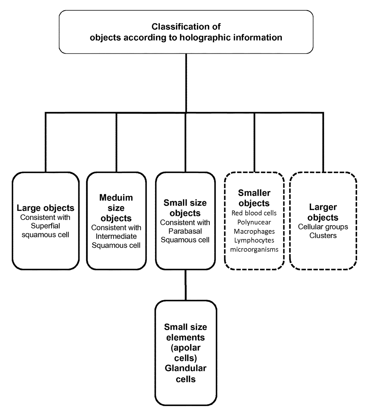
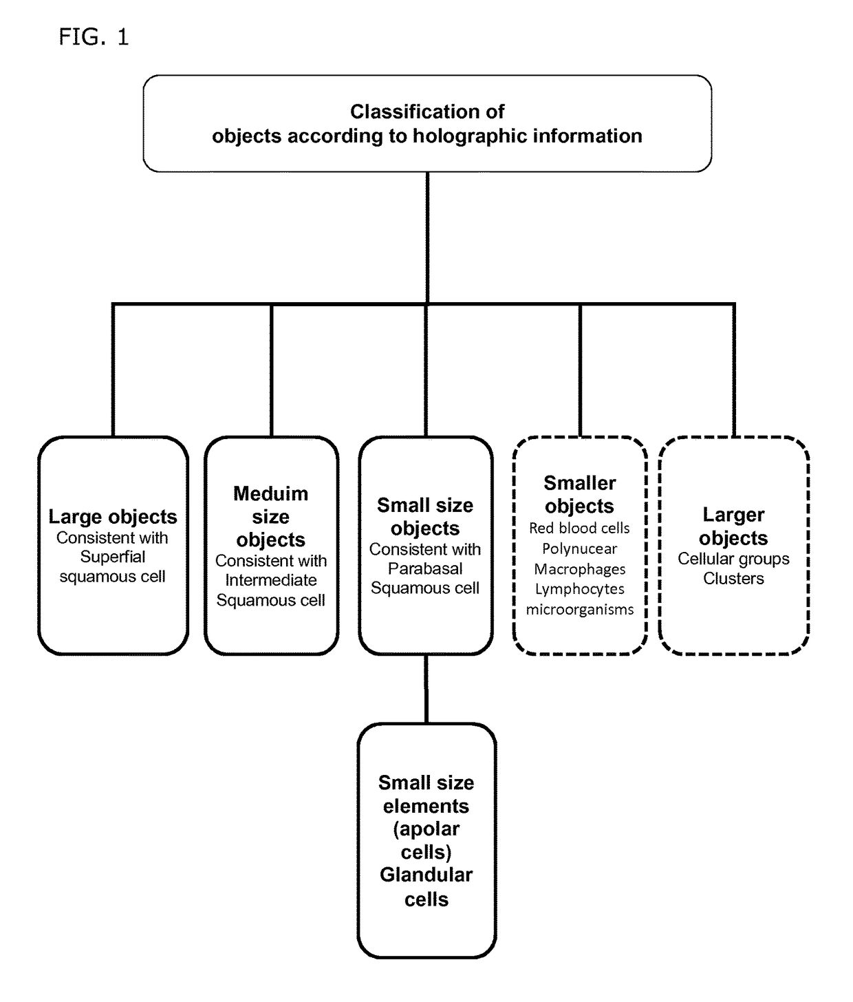
[0143]16 selected patients previously diagnosed by the Thinprep® liquid based cytology confirmed by HPV Abott® assay or histology diagnosis for CIN2 / 3, were analysed on the new Holocyt® diagnostic intelligence software by use of the Holographic Digital Microscope (DHM) using partially coherent laser light.

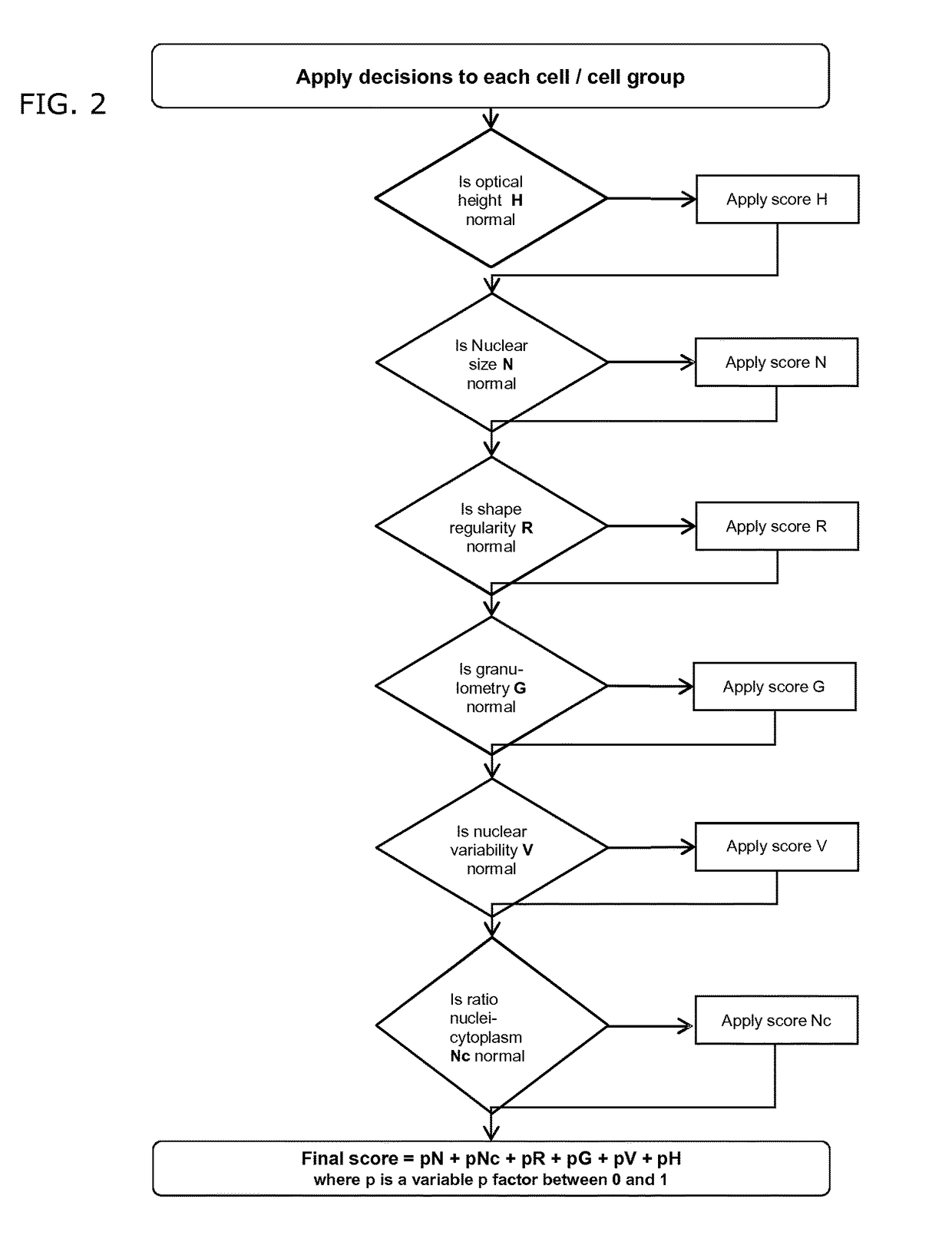
[0144]DHM enables a quantitative multifocal phase contrast imaging that has been found suitable for quantitative and qualitative inspection, and for 3-dimensional cell imaging. 188 cells were identified and measured in an automated way. Nucleus / Cell Ratio (NCR) and Optical Height Delta (OHD) were extracted in the 3D holographic image. The Optical Height Delta is the difference between Nucleus top height minus Cytoplasm average height. NCR and OHD were separately determined in 2 groups: CIN1 or CIN 2 / 3 patients.

[0145]These results were compared with normal cells either from patients with normal cytology diagnosis either from normal cells within the abnormal smears. Data were importe...


PUM

PropertyMeasurementUnit
cell sizeaaaaaaaaaa
cell sizeaaaaaaaaaa
cell sizeaaaaaaaaaa

Abstract

The current invention concerns a method for detecting cancerous cells and / or classifying cells in a cell sample comprising the following steps: providing a cell sample; obtaining holographic information from said cell sample by digital holographic microscopy (DHM); deriving at least one cellular parameter from said holographic information, and; classifying said cells of cells sample; characterized in that said classification occurs by appointing a Scoring Factor to said cells of cell sample, based on said cellular parameters. In a second aspect, a system for the detection of cancerous cells and / or classification of cells in a cell sample is provided, employing the method as disclosed in the invention. In a final aspect, a method for updating and / or improving a database comprising thresholds linked to holographic information and the database related thereof is equally disclosed.

Description

CROSS-REFERENCE TO RELATED APPLICATIONS[0001]This application is the U.S. National Phase under 35 U.S.C. §371 of International Application PCT / EP2012 / 063936, filed Jul. 16, 2012, which claims priority to EP 11174583.2, filed Jul. 19, 2011 and PCT / EP2011 / 070571, filed Nov. 21, 2011.TECHNICAL FIELD[0002]The invention pertains to the technical field of diagnostics, and more specifically to a method and system for determining specific parameters of a cell sample by obtaining holographic information by digital holographic microscopy. The method offers a non-destructive manner of analyzing cells and can be used for the detection of cancerous cells and classification of the cells present in a cell sample. Holographic information is obtained and compared to a threshold database relating to cellular parameters. The present invention furthermore discloses a system that employs the method by digital holographic microscopy of the invention as well as a method for updating and / or improving a dat...

Claims


Application Information

Patent Timeline
Patent Type & Authority Patents(United States)
IPC IPC(8): G06K9/62G02B21/00G06K9/00G03H1/00G03H1/04G02B21/36G01N21/45G01N33/483G16B20/00G16B50/00
CPCG03H1/0443G01N21/45G01N33/4833G02B21/0004G02B21/365G03H1/0005G06K9/00134G03H2001/005G03H2210/12G06F16/51G16B20/00G16B50/00G06V20/693G06F16/00G01B9/021G01N15/14G01N33/483G01N33/574G06F18/00G06V20/698G01N15/1434G01N15/1475G03H2001/0452G03H2226/02
Inventor MATHUIS, PHILIPJOORIS, SERGEMAGNIETTE, OLIVIER
Owner OVIZIO IMAGING SYST NV SA
Why Eureka
Try Eureka