

Collagen scaffolds
a scaffold and collagen technology, applied in the field of collagen scaffolds, can solve the problems of premature loss of fibrin clot scaffold, tissue that fails to heal after an injury, and disrupts the healing process of tissues within the joint or intra-articular tissues,
- Summary
- Abstract
- Description
- Claims
- Application Information
AI Technical Summary
Benefits of technology
Problems solved by technology
Method used
Image

Examples
example 1
Terminal Sterilization Study
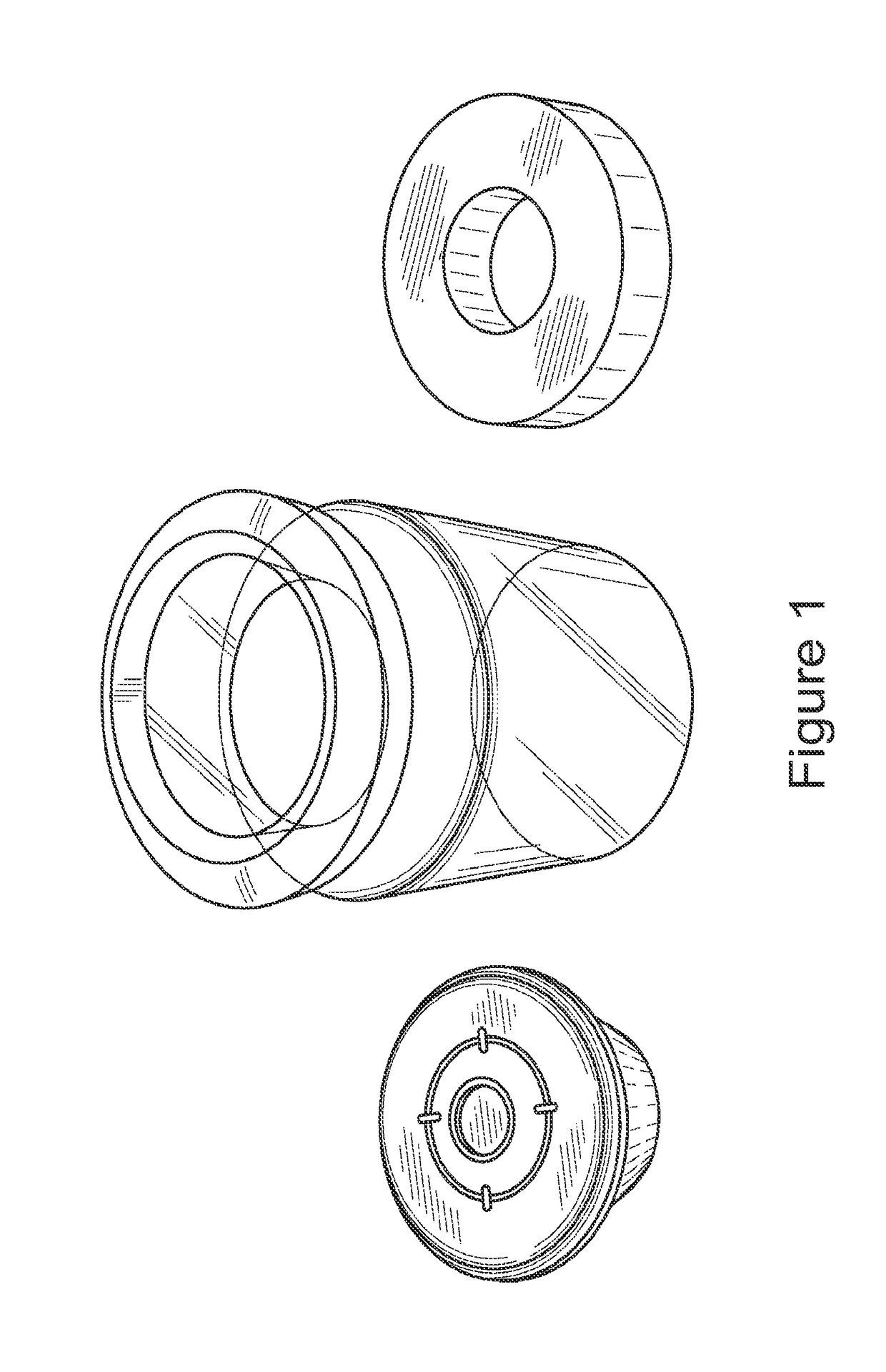
[0157]Methods: In this study, an extracellular matrix (ECM) scaffold prepared according to the methods of the invention was terminally sterilized using standard E-beam and EO sterilization. Briefly, the scaffolds were prepared by lyophilizing bovine connective tissue, washing the tissue, digesting in pepsin and then lyophilizing in a mold to make a cylindrical scaffold. The samples were placed inside glass vials. The glass vials were purged by Nitrogen in order to reduce the amount of the oxygen in them. The glass vials were then sealed and clamped as shown in FIG. 1 and stored on dry ice throughout the sterilization procedure.
[0158]E-Beam: For the e-beam groups, the ECM scaffolds were placed in glass vials that were capped and sealed using plastic stoppers and crimped using an aluminum clamping system. The samples were then subjected to 15 kGy E-Beam sterilization process by Sterigenics (San Diego, Calif.). An “e-beam” control group of scaffolds was ship...
example 2
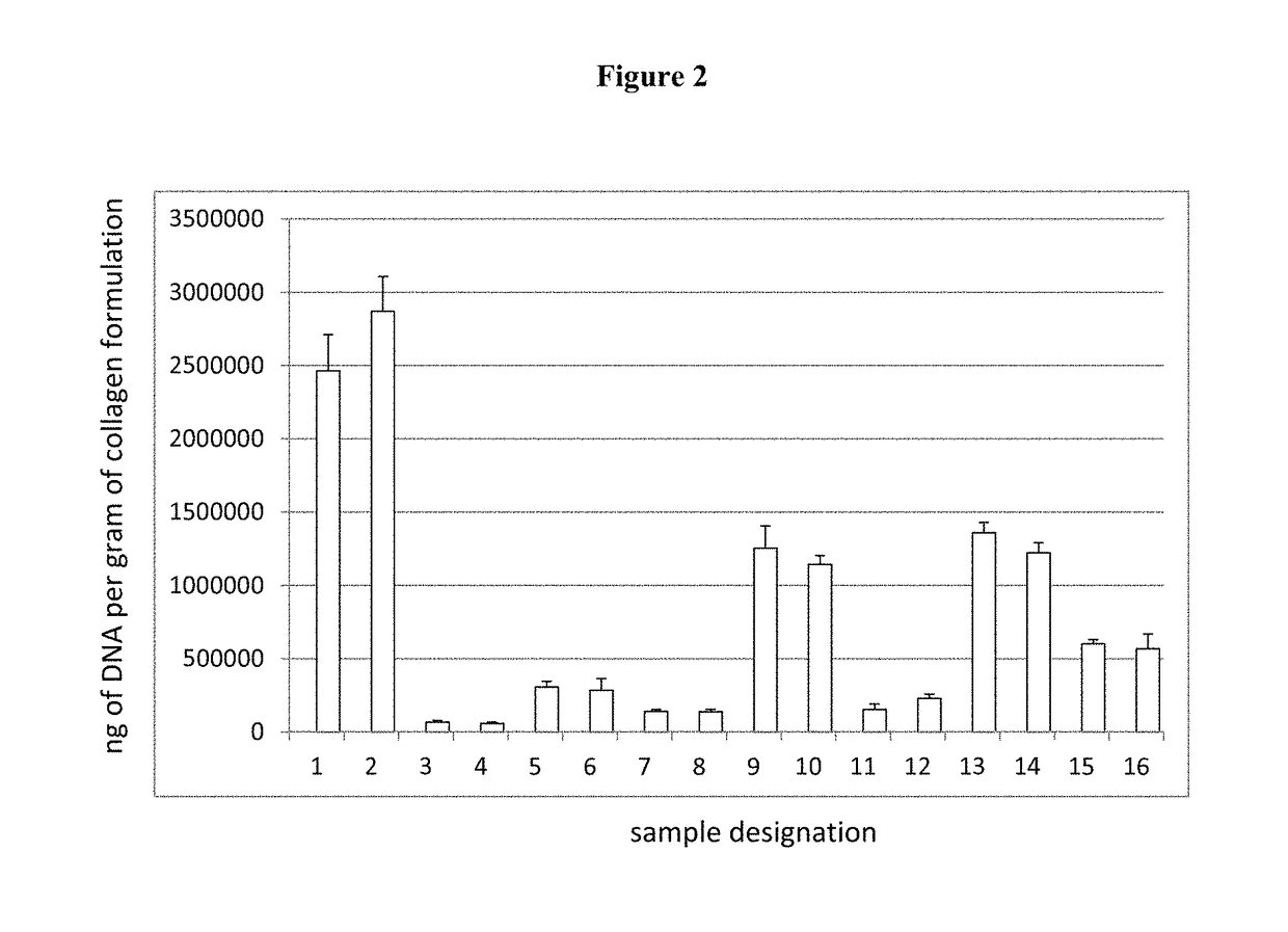
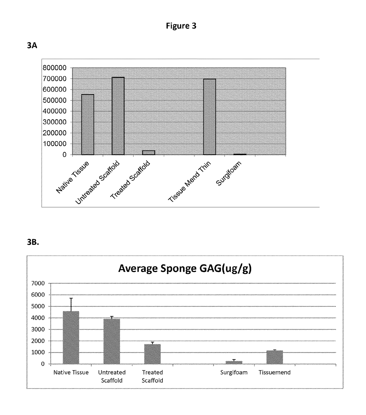
Evaluation of DNA Content / RNA Content / Cell Fragment Content in Scaffolds
[0180]Scaffold were treated with various chemicals and enzymes to lower the DNA, GAG and Phospholipid content. As described in detail below it was possible to significantly reduce the DNA, GAG and Phospholipid content of the scaffolds.
[0181]In this experiment, tissues from 8 bovine knees were collected. The total wet weight of the harvested tissues was 166.5 g. The tissues were lyophilized until dry and then homogenized. The homogenized tissue was then divided into 16 samples with dry weights.
[0182]The dry tissue samples were then rinsed in 2% antibiotic solution overnight.
Start of Differentiation:
Following the antibiotic rinse, the samples were divided into 16 different treatment groups as noted below.
Summary of Sample Groups: Differences Only are Noted in Table 2.
[0183]Sample 1: The tissue was rinsed with NaCl solution followed by ultrapure water three times, and then washed with citrate buffer with a pH=4.0 f...
example 3
Chemical Neutralization of the Pepsin Content in a Scaffold
[0204]Scaffolds were made from extracellular matrix proteins using a pepsin digestion. After digestion, one group had no further treatment, while the second group was treated with a chemical to inactivate the pepsin.
[0205]Scaffolds were made from extracellular matrix proteins using a pepsin digestion. After digestion, one group had no further treatment, while the second group was treated with a strong base to inactivate the pepsin.
[0206]Briefly, a strong base (e.g., KOH or NaOH or LiOH) at a suitable concentration was added into a collagen slurry in a dropwise manner to bring the pH value to above 4.0. Additional strong base was added to bring the pH of the slurry to 7.0 or greater. Once the slurry reached its target pH range, the solution is kept there for a suitable period of time, e.g., 1 to 10 minutes.
[0207]After inactivation of the pepsin, the pH of the slurry was returned to a pH between 7.0 and 8.0 by the addition of ...
PUM
| Property | Measurement | Unit |
|---|---|---|
| concentration | aaaaa | aaaaa |
| time | aaaaa | aaaaa |
| temperature | aaaaa | aaaaa |
Abstract
Description
Claims
Application Information