In vitro three-dimensional culture model of glioma stem cells and application thereof

A technology of glioma stem cells and glioma cells, which is applied in the field of cell biology, can solve problems such as cell damage, Matrigel glue is not easy to dissolve, and limits the research progress of vascular mimicry in vitro, and achieves the effect of convenient use and simple operation

Active Publication Date: 2015-01-28
THE FIRST AFFILIATED HOSPITAL OF THIRD MILITARY MEDICAL UNIVERSITY OF PLA
View PDF1 Cites 9 Cited by
  • Summary
  • Abstract
  • Description
  • Claims
  • Application Information

AI Technical Summary

Problems solved by technology

However, Matrigel glue is essentially a kind of Matrigel, which only provides a matrix for cell culture, not a real three-dimensional culture system.
In addition, due to the reasons of Matrigel itself, the following disadvantages were found during the research process: (1) Matrigel is jelly-like at room temperature, and the operation must be carried out in a low temperature environment below 4°C, which will cause damage to cells; (2) Because some cell phenotypes of cells in two-dimensional cu

Method used


Image

  • In vitro three-dimensional culture model of glioma stem cells and application thereof
  • In vitro three-dimensional culture model of glioma stem cells and application thereof
  • In vitro three-dimensional culture model of glioma stem cells and application thereof

Examples


Embodiment 1

[0027] Embodiment 1 GFP-U87 and GFP-091214 sphere-forming experiment

[0028]In a 100mm petri dish, the green fluorescently labeled U87 (GFP-U87) was cultured in DMEM medium containing 10% fetal bovine serum (Gibco, 10099-133) for 3 days, and the medium in the petri dish was aspirated and discarded. Add 5ml of PBS to wash, discard, and repeat twice; add 10ml of stem cell culture medium, and then add new stem cell culture medium in a half-change method every day, and the balls can be formed in 5-7 days, and the balls of GFP-U87 can be obtained. For breeding and experimentation.

[0029] Green fluorescently labeled primary glioma cells 091214 (GFP-091214) were cultured according to the above method to obtain spherical GFP-091214 for passage and experiment.

Embodiment 2

[0030] Example 2 Establishment of an in vitro three-dimensional culture model of glioma stem cells

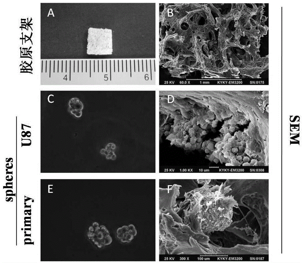
[0031] Soak the three-dimensional collagen scaffold in DMEM overnight, remove the excess DMEM adhered to the scaffold with filter paper, place the scaffold in a six-well plate, and plant the sphered GFP-U87 and GFP-091214 cells in Example 1 respectively Put it on the collagen support and let it stand in an incubator at 37°C for 4 hours, then add 5ml of stem cell culture medium to culture for 3 days, and use the GFP-U87 and GFP-091214 cell spheres cultured in the culture dish of Example 1 as a control, and observe the results with a scanning electron microscope showed that glioma stem cells grow in a three-dimensional manner within the collagen scaffold, such as figure 1 D and figure 1 As shown in F, the structure of the GFP-U87 and GFP-091214 cell spheres cultured in the culture dish is as follows figure 1 C and figure 1 Shown in E.

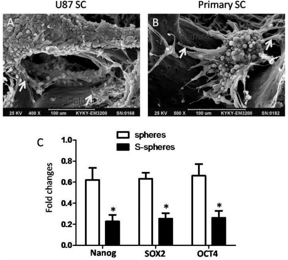
[0032] RT-PCR was used to detect the ex...

Embodiment 3 3

[0033] The comparison of embodiment 3 three-dimensional collagen support and Matrigel glue culture model

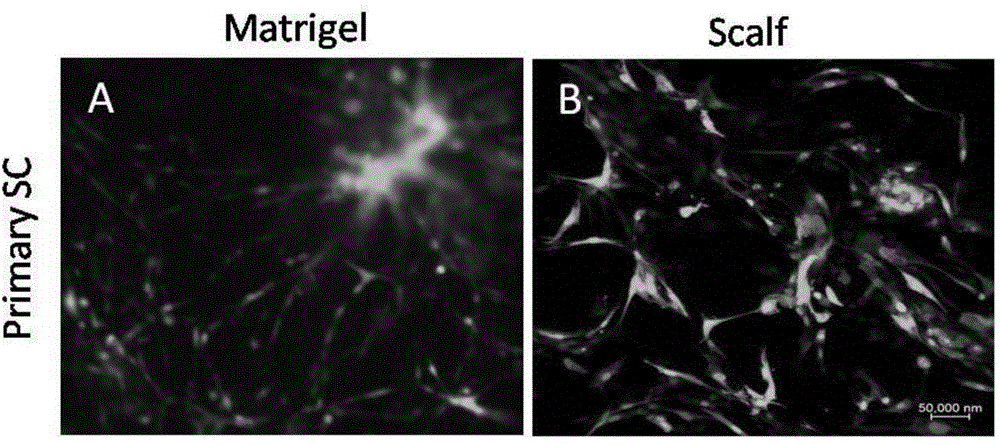
[0034] Soak the three-dimensional collagen scaffold in DMEM overnight, remove the excess DMEM adhered to the scaffold with filter paper, place the scaffold in a six-well plate, and inoculate the GFP-091214 cell spheres formed in Example 1 on the three-dimensional collagen scaffold and Matrigel glue, after 4 hours in a 37°C incubator and cultured in endothelial medium for 3 days, confocal laser microscopy showed that glioma stem cells formed tube-like structures on both Matrigel glue and three-dimensional scaffolds. In the three-dimensional collagen scaffold, it can be seen that the cells form a three-dimensional tube-like structure, while the tube-like structure formed by Matrigel glue is mainly formed by cell protrusions. The results are as follows image 3 shown.


PUM


Abstract

The invention belongs to the field of cytobiology and specifically relates to an in vitro three-dimensional culture model for research on vasculogenic mimicry of glioma stem cells and an application thereof. Preparation of the in vitro three-dimensional culture model comprises the following steps: immersing a three-dimensional collagen scaffold in a DMEM culture medium for 6-24 h, taking out the three-dimensional collagen scaffold, removing superfluous DMEM adhered to the three-dimensional collagen scaffold to obtain a pretreated three-dimensional collagen scaffold, placing the pretreated three-dimensional collagen scaffold on a cell culture plate, culturing sphere-formed glioma stem cells in the pretreated three-dimensional collagen scaffold, standing at 37 DEG C for 2-6 h, adding an endothelial medium, and culturing at 37 DEG C for 2-4 days, so as to obtain the in vitro three-dimensional culture model for research on vasculogenic mimicry of glioma stem cells. Each operational process of the in vitro three-dimensional culture model is carried out at room temperature; various kinds of in situ staining, such as immunohistochemical staining and immunofluorescent staining, can be carried out; electron microscopic examination can be carried out; and RT-PCR and Western Blot detection can be carried out directly after digestion.

Description

technical field [0001] The invention belongs to the field of cell biology, and in particular relates to an in vitro three-dimensional culture model for studying vascular mimicry of glioma stem cells and its application. Background technique [0002] The growth of tumors and the formation of distant lesions depend on blood supply. According to the classic theory of angiogenesis, when a solid tumor grows to 1-2mm 3 When tumors are vaccinated, they need to induce the formation of new blood vessels to obtain blood supply, otherwise the tumor will stop growing. With the deepening of the research on tumor biology, in recent years, people have also discovered that tumor cells can deform themselves and form a lumen around them, in a way that is independent of endothelial cell-dependent vascular mimicry (VM). Construct tumor microcirculation. Since Maniotis et al. reported the existence of a new tumor microcirculation model, vascular mimicry (VM), in malignant melanoma in 1999, peo...

Claims


Application Information

Patent Timeline
IPC IPC(8): C12N5/095G01N33/68
Inventor 姚小红吴海波卞修武戴建武
Owner THE FIRST AFFILIATED HOSPITAL OF THIRD MILITARY MEDICAL UNIVERSITY OF PLA
Why Eureka
Try Eureka