Imaging individual mRNA molecules using multiple singly labeled probes

a single labeled probe and mrna technology, applied in the direction of microbiological testing/measurement, biochemistry apparatus and processes, etc., can solve the problems of inability to precisely determine the location of the target molecule, poor sensitivity, and inability to widely adopt a system

Active Publication Date: 2018-02-20
RUTGERS THE STATE UNIV
View PDF16 Cites 15 Cited by
  • Summary
  • Abstract
  • Description
  • Claims
  • Application Information

AI Technical Summary

Benefits of technology

[0015]A computer program can be used to identify and count all the mRNA molecules in the cell from the microscopic image. In situ hybridizations performed with the sets of probes described above allow accurate and simple gene expression analysis, detection of pathogens and pathogenic states such as cancer.

Problems solved by technology

However, because the products of these reactions were small molecules or precipitates that diffuse away from the probe, the location of the target molecules could not be precisely determined.
Conversely, probes labeled directly with a few fluorophores maintained spatial resolution, but the sensitivity that can be achieved is relatively poor.
Although the authors convincingly demonstrated single molecule sensitivity and other groups have successfully used these probes, the system has not been widely adopted.
One reason for this is difficulty in the synthesis and purification of heavily labeled oligonucleotides.
When multiple amino groups are introduced into the same oligonucleotide some are lost due to side reactions such as transamidation.
Coupling of fluorophores to the remaining amino groups is inefficient and requires several consecutive coupling reactions and it is difficult to purify oligonucleotides in which all designed sites are coupled to fluorophores from those that are partially coupled.
Also, when some fluorophores are present in multiple copies on the same oligonucleotide they interact with each other altering the hybridization characteristics of the oligonucleotides and exhibiting severe self-quenching.
Another issue with the use of small numbers of heavily labeled probes is that a significant portion of the fluorescence is lost for every probe that does not bind to the target, whereas every non-specific binding event increases the background.
This makes it difficult to unambiguously identify those fluorescent spots as mRNA molecules, since it is impossible to determine whether the detection of an individual probe arises from legitimate binding to the target mRNA or non-specific binding.
These “thresholding” problems limit the ability of such methods to provide reliable counts of mRNA numbers in individual cells.

Method used


Image

  • Imaging individual mRNA molecules using multiple singly labeled probes
  • Imaging individual mRNA molecules using multiple singly labeled probes
  • Imaging individual mRNA molecules using multiple singly labeled probes

Examples


example 1

Materials and Methods

[0056]The procedures described in this section are applicable to all examples unless indicated otherwise.

[0057]Probe Design

[0058]The sets of probes were designed to consist of at least 48 oligonucleotides each with lengths varying from 17 to 22 nucleotides long with a 3′-amine modification (FKBP5, FLJ11127, and Map2 mRNAs were probed using 63, 53 and 72 oligonucleotides respectively). Additionally, the GC content of the oligonucleotides was kept close to 45% when possible. The oligonucleotides were pooled and coupled to a fluorophore in a single reaction, after which the uncoupled oligonucleotides and remaining free fluorophores were removed by HPLC purification.

[0059]Fluorescence in situ Hybridization

[0060]In preparation for FISH, all samples were fixed with 3.7% formaldehyde and permeabilized with ethanol. The hybridization was performed using buffers and conditions similar to those outlined by Femino et al., with the key difference being the stringency of the...

example 2

Probing Repeated and Unique Sequences Present in the Same mRNA Molecule

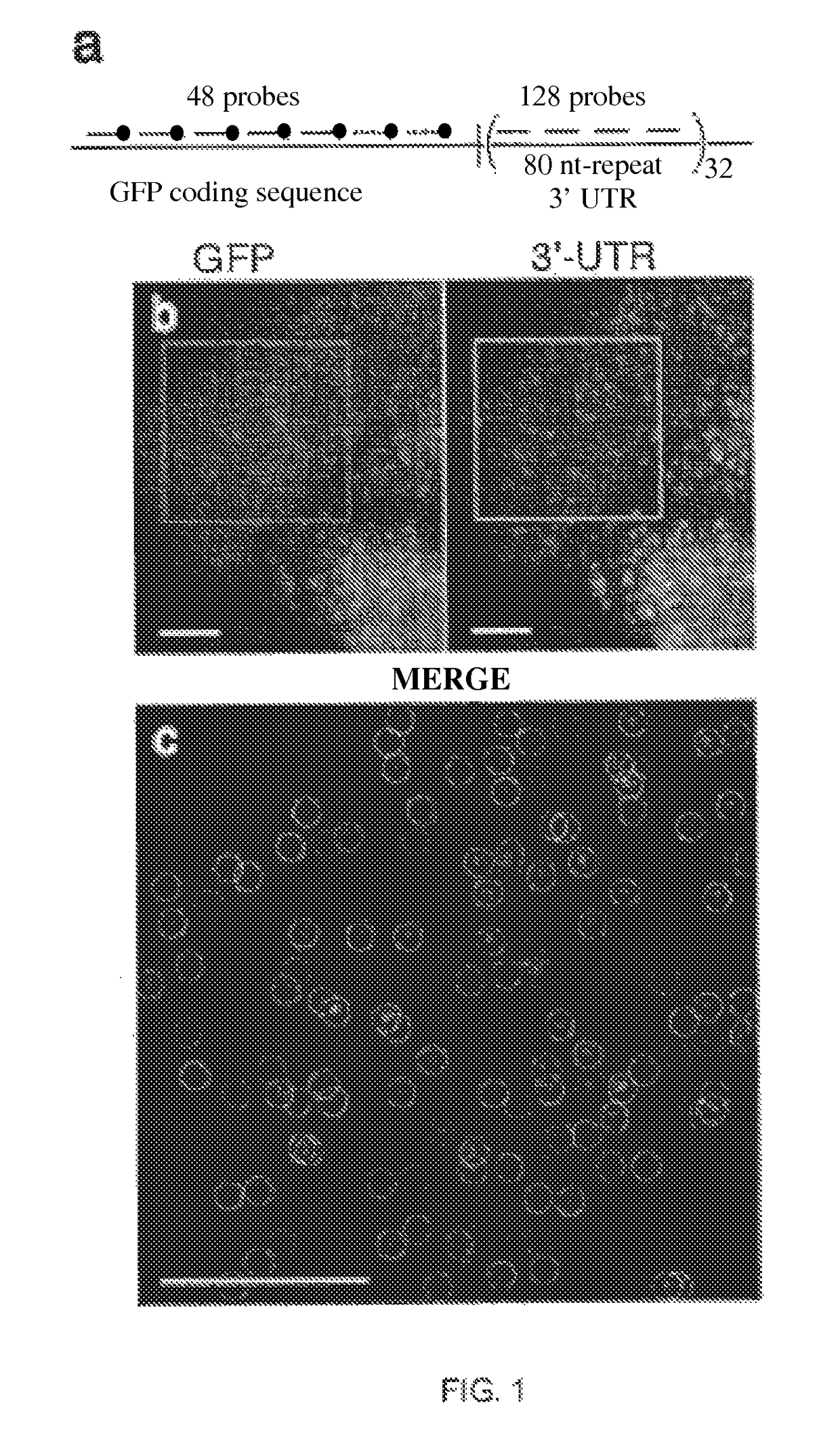
[0063]Utilizing small oligonucleotide probes labeled with a single fluorophore moiety, the inventors have shown that individual mRNA molecules that were engineered to contain 32-96 tandem copies of a probe-binding sequence can be detected by in situ hybridization. The inventors also demonstrated that the individual spots in the image represent single mRNA molecules, utilizing a number of different approaches, including correlating the average mRNA copy number obtained by directly counting the diffraction-limited spots to a measurement of the number of target molecules obtained by real-time RT-PCR. Thus, if many different probes are utilized, each targeted to a distinct region of a natural mRNA, it would be possible to obtain single-molecule sensitivity without resorting to the use of engineered genes.

[0064]For the initial test of this hypothesis, the inventors constructed a doxycycline-controlled gene that produc...

example 3

Computational Algorithm for Spot Detection

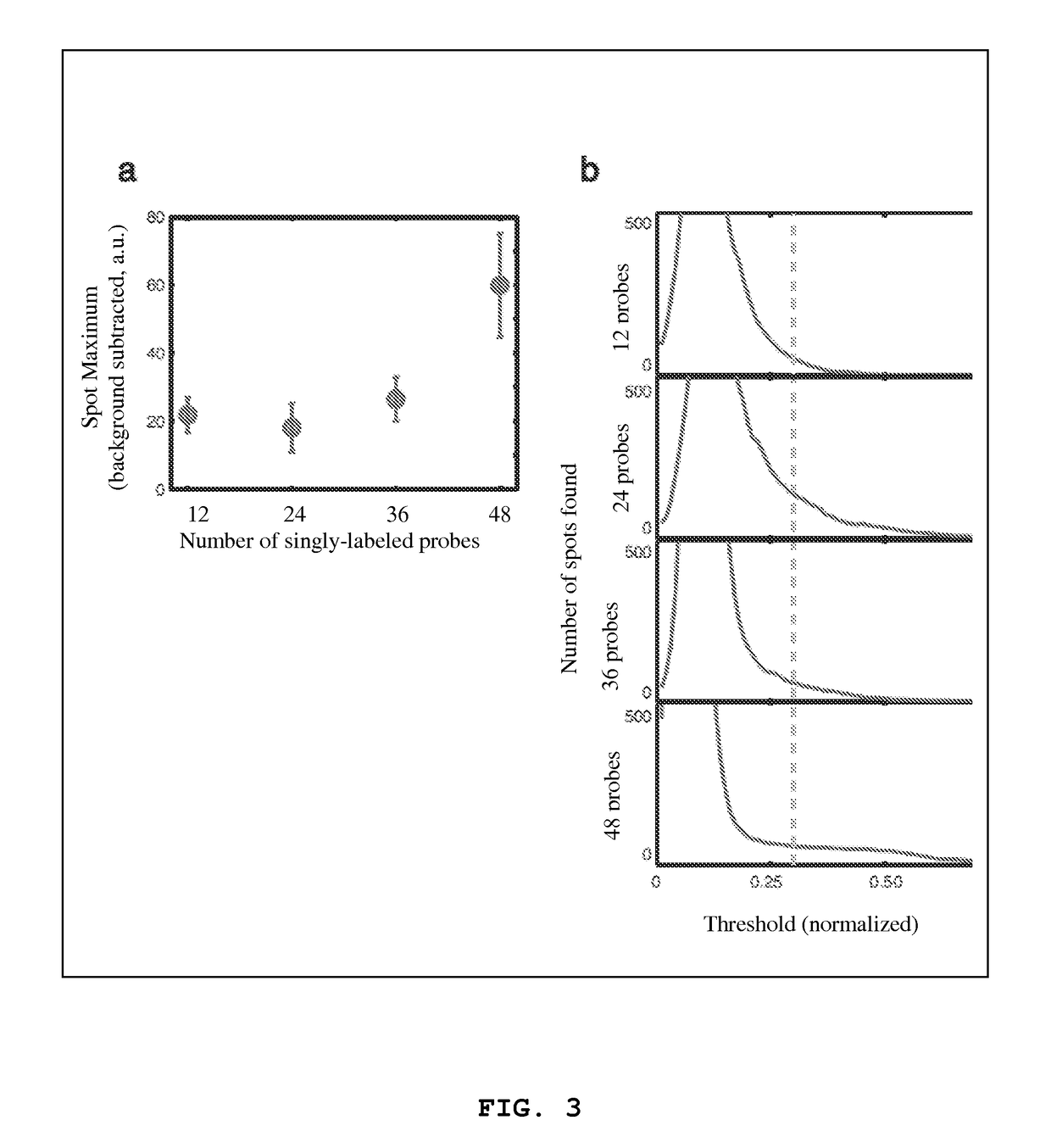
[0069]In order to reliably identify large numbers of mRNA molecules, the inventors developed a semiautomated computational algorithm for finding spots in a three-dimensional stack of fluorescent images. One of the difficulties associated with spot detection is the nonuniform background arising from cellular autofluoresence and low levels of non-specific probe hybridization. To circumvent these issues, the inventors filtered image stacks using a three dimensional linear Laplacian of Gaussian filter designed to enhance spot-like signals of the correct size and shape (FIG. 5A and FIG. 5B) while removing the slowly varying background. In the next step in the algorithm, the inventors applied a threshold to the filtered image in order to define the spots. In order to make a rational choice of threshold, the number of spots in three dimensions for all thresholds ranging from zero to the maximum pixel intensity in the filtered image was counted. Whe...


PUM

PropertyMeasurementUnit
diameteraaaaaaaaaa
binding affinityaaaaaaaaaa
fluorescentaaaaaaaaaa

Abstract

A method for probing a target sequence of messenger ribonucleic acid molecules (mRNA's) in a fixed, permeabilized cell, said target sequence including at least 30 non-overlapping probe binding regions of 15-100 nucleotides, comprising immersing said cell in an excess of at least 30 nucleic acid hybridization probes, each singly labeled with the same fluorescent label and each containing a nucleic acid sequence that is complementary to a different probe binding region of said target sequence; washing said fixed cell to remove unbound probes; and detecting fluorescence from said probes.

Description

RELATIONSHIP TO PRIOR APPLICATIONS[0001]This application is the U.S. National Phase of International Patent Application Ser. No. PCT / US09 / 56564, filed Sep. 10, 2009, which claims priority to a U.S. Provisional Patent Application Ser. No. 61 / 191,724, filed on Sep. 10, 2008, both of which are incorporated herein by reference in their entireties.GOVERNMENT INTERESTS[0002]The invention disclosed herein was made, at least in part, with Government support under Grant Nos. NIH GM-070357 and NIH MH-079197 from the National Institutes of Health. Accordingly, the U.S. Government has certain rights in this invention.FIELD OF THE INVENTION[0003]This invention generally relates to methods of nucleic acid sequence detection.BACKGROUND[0004]As it has become increasingly apparent that gene expression in individual cells deviates significantly from the average behavior of cell populations, new methods that provide accurate integer counts of mRNA copy numbers in individual cells are needed. Ideally, ...

Claims


Application Information

Patent Timeline
Patent Type & Authority Patents(United States)
IPC IPC(8): C12Q1/68
CPCC12Q1/6827C12Q1/6841C12Q2537/143C12Q2565/102
Inventor RAJ, ARJUNTYAGI, SANJAY
Owner RUTGERS THE STATE UNIV
Why Eureka
Try Eureka