Heterogenous skin transfected by recombined genes

A skin and wound technology, applied in the field of isolated pig skin, can solve problems such as difficulty, no application, limited effect, etc., achieve good growth conditions, reduce wound scar formation, and promote healing

Inactive Publication Date: 2008-05-14
CHONGQING ZONGSHEN JUNHUI BIOTECH
0 Cites 0 Cited by

AI-Extracted Technical Summary

Problems solved by technology

In the prior art, there is no application of it in burn wound skin grafting
At the same time, due to the difficulty in the preparation and purification of CTLA4, the cost of systemic or even local application is expensive, and studies have foun...

Abstract

A heterogenous skin transfected by recombined gene CTLA4Ig and able to express the recombined protein CTLA4Ig is disclosed. This invented heterogenous skin can be used to cover and seal the burn wound, promote wound heeling and reduce the scar formation.

Application Domain

Technology Topic

Burn woundMedicine +2

Image

  • Heterogenous skin transfected by recombined genes
  • Heterogenous skin transfected by recombined genes
  • Heterogenous skin transfected by recombined genes

Examples

  • Experimental program(6)

Example Embodiment

[0021] Example 1
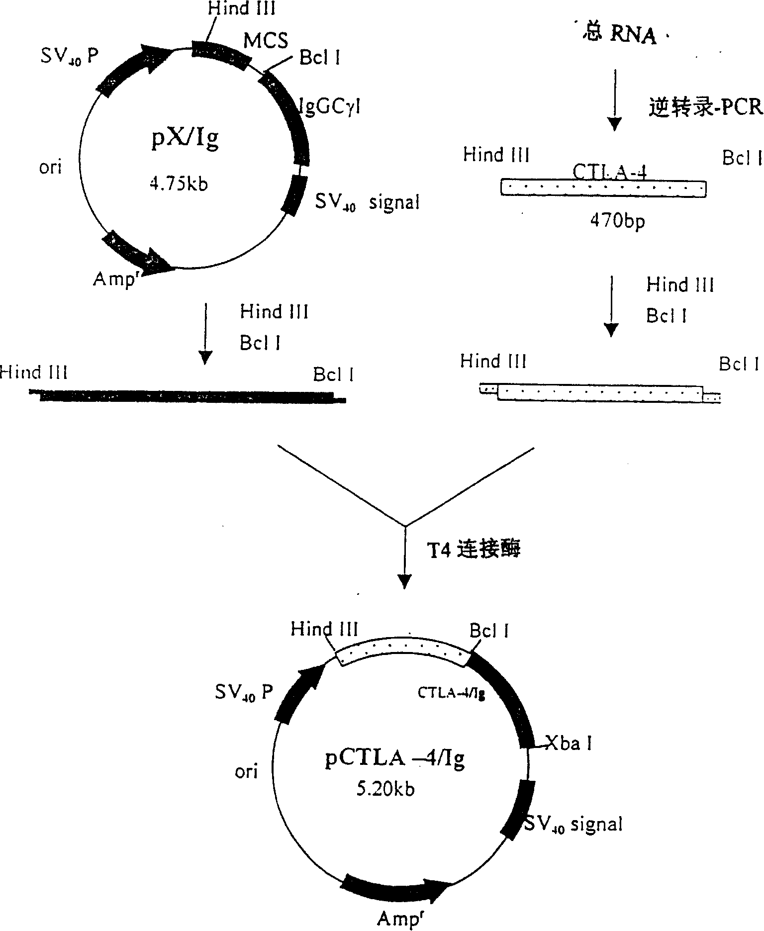
[0022] Construction of Ig fusion protein expression vector ( figure 1 )
[0023] 1. Separation and activation of peripheral blood mononuclear cells (PBMC)
[0024] Routinely isolate mononuclear cells from healthy human peripheral blood, RPMI1640 adjusts the cell density to 1×10 6 Cells/ml, add PMA (15nM) and anti-CD3 monoclonal antibody (10μg/ml), 37℃ 5% CO 2 Cultivate for 6 hours to activate lymphocytes and highly express CTLA-4mRNA.
[0025] 2. Extraction and identification of total cell RNA
[0026] 1) Extraction: collect activated PBMC by centrifugation, wash the cells with PBS (DEPC) × 2, press each 1 × 10 7 Add 1 ml of Tripure to PBMC, mix well, RT for 5 minutes, add 0.2 ml of chloroform, shake vigorously for 15 seconds, RT for 5 minutes, 10,000 rpm/centrifuge for 15 minutes, transfer the upper colorless water phase to another centrifuge tube, add Mix 0.5 ml of isopropanol, -20°C for 10 minutes, 10,000 rpm/centre for 15 minutes, add 1 ml of 75% ethanol to wash the precipitate, add 200 μl DEPC water to dissolve the precipitate after it is dry, and store at -20°C.
[0027]2) RNA electrophoresis: 1.5% formaldehyde denatured agarose gel to the plate to make the gel, and put it in 1×MOPS electrophoresis solution after coagulation; 5 microliters of RNA sample and 15 microliters of loading buffer, denaturation in 95℃ water bath for 2 minutes, 50V , Electrophoresis, UV detection of RNA integrity.
[0028] 3) RNA content determination: the RNA sample is diluted and measured O.D. on the UV spectrophotometer. 260 And O.D. 280 Value, RNA concentration (μg/ml) = O.D. 260 ×40×dilution factor.
[0029] 3. Primer design
[0030] Through computer-aided analysis, according to the human CTLA-4 gene sequence in the gene bank, the following three primers were designed, among which primer 1# includes 7 amino acid residues at the N-terminal of CTLA-4 extracellular region and 15 C-terminals of the oncostatin M signal peptide Amino acid residues; Primer 2# corresponds to CTLA-4119-125 amino acid residues, and introduces a Bcl I restriction site; Primer 3# contains the remaining amino acid residues of the oncostatin M signal peptide and is combined with primer 1# for tumor suppression The prime M signal peptide partially overlaps, and a HindIII restriction site is introduced at the 5'end of primer 3#. The primers were synthesized by Shanghai Institute of Cell, Chinese Academy of Sciences and purified by PAGE.
[0031] Primer 1#: CTCAGTCTGGTCCTTGACCTCCTGTTTCCAAGCATGGCGA
[0032] GCATGGCAATGCACGTGGCCCAGCC
[0033] Primer 2#: TTTGGGCTCCTGATCAGAATCTGGGCACGGTTC
[0034] ——Bcl I
[0035] Primer 3#: GCAAGCTCAATGGGTTGACTGCTCACACAGAGGACGCTG
[0036] ——HindIII
[0037] CTCAGTTCGGTCCTTGCATCT
[0038] 4. RT-PCR amplification of CTLA-4 extracellular region cDNA fragment
[0039] 1) cDNA first strand synthesis (reverse transcription): 10 microliters of total RNA, oligo(dT 12 ) 2 microliters, ddH 2 O(DEPC) 11.5 microliters, after mixing, denature in 65℃ water bath for 10 minutes, quickly place on ice, then add 8 microliters of 5× reverse transcription buffer, 100mM DTT 2 microliters, RNasin 0.5 microliters, 2mM dNTP 4 Microliter, 2 microliters of AMV, (total volume 40 microliters), 42°C for 1 hour, store at -20°C.
[0040] 2) PCR reaction: firstly use the first strand of cDNA as a template, and use primers 1# and 2# for the first round of amplification. The conditions are: 5 microliters of the first strand of cDNA, 5 microliters of 10×PCR buffer, 10mM dNTP4 Microliter, primer 2# and 3# each -1 microliter (final concentration 100nM), ddH 2 O 29.5 μl, denaturation at 95°C for 7 minutes, add 0.5 μl Taq enzyme, total volume 50 μl, cycle conditions: 94°C for 1 minute, 55°C for 1 minute, 72°C for 1 minute, 72°C for 7 minutes after 30 cycles ; Then use the first round of PCR reaction product as a template and primers 2# and 3# for the second round of amplification. The reaction conditions are the same as before, and the amplified products are detected by 2% agarose gel electrophoresis.
[0041] 5. Plasmid extraction
[0042] Operate according to the instructions of the plasmid extraction kit, 100 microliters of ddH 2 O, 1 eluted plasmid DNA.
[0043] 6. E. coli competent, cryopreserved
[0044] Refer to "Molecular Cloning" (Jin Dongyan, Li Mengfeng translation, "Molecular Cloning Experiment Guide", second edition Beijing: Science Press 1996; P852-907) CaCl 2 Prepare competent E. coli, add 10-15% glycerol to final concentration, and freeze at -70°C.
[0045] 7. Transformation of plasmid and recombinant DNA
[0046] Take the cryopreserved E. coli competent bacteria, add 1-2 microliters of plasmid or DNA ligation product, mix gently, ice bath for 30 minutes, 42°C water bath for 2 minutes, add 900 microliters of LB medium, 37°C water bath 60 Take an appropriate amount and spread it on an LB agar plate containing 100 μg/ml ampicillin. Cultivate overnight at 37°C incubator.
[0047] 8. Recovery of DNA fragments
[0048] Follow the instructions of the DNA fragment recovery kit.
[0049] 9. Connection between PCR amplified fragments and Ig fusion protein expression vector
[0050] The second round of PCR amplification products were subjected to 2% agarose gel electrophoresis, and the required fragments were recovered. After double digestion with HindIII and Bcl I, the fragments of pX/Ig vector (gifted by others) were digested with the same enzymes: vector 3 :1 ratio, ligate according to the instructions of the DNA rapid ligation kit, transform MC1061 (ATCC, USA) competent bacteria, HindIII, Xba I enzyme digestion to identify the insert size of the positive transformed clone.
[0051] 10. Sequence determination
[0052] Recombinant plasmids with the correct insert size were identified by restriction enzyme digestion. After digestion with HindIII and Xba I, they were inserted into pUC19 plasmid (TaKaRa, Japan). Sanger dideoxy sequencing method was used. The plasmid double-stranded DNA was used as the template and the universal primers were used in pUC19. Under the guidance, the ABI Prism377 automatic sequencer automatically sequenced.

Example Embodiment

[0053] Example 2 Construction of CTLA-4Ig retroviral expression vector
[0054] The pLXSN (Clontech, USA) and pUC19-CTLA-4Ig were digested with EcoRI and HindIII respectively, and then filled with dATP, dNTP and DNA E. coli polymerase Klenow fragment respectively, and then digested with BamHI, and electrophoresed The large fragment of pLXSN is recovered and the small fragment of pUC19-CTLA-4Ig is connected to obtain the expression pLXCTLA-4Ig of CTLA-4Ig.

Example Embodiment

[0055] Example 3 Construction of CTLA4Ig adenovirus expression vector
[0056] 1. Blunt the ends of the cDNA fragments.
[0057] Add 1μl of 10× buffer solution to the alcohol precipitation cDNA (0.5pmol) centrifuge tube, then add sterile double distilled water to 9μl, 70℃ for 5 minutes, add 1μl T4DNA polymerase, mix gently, do not shake vigorously, incubate at 37℃5 After 10 minutes, adjust the DNA concentration to 1μg and 50μl with DNA Dilition buffer, and mix well. Transfer to an ice bath to inactivate the polymerase, which can be used directly in the ligation reaction (if it can not be used immediately, it should be extracted and precipitated immediately, dissolved in DNA Dilition buffer and stored at -20°C).
[0058] 2. Insert the blunt-ended cDNA into pAxCAwt (TakaRa, Japan) Korotkoff plasmid.
[0059] Linearize pAxCAwt: pAxCAwt 5μl, h buffer (high salt buffer) 5μl, Swa I 2μl, ddH 2 O 38μl, mix well and incubate at 25°C for 2 hours; add EDTA solution to a final concentration of 10mM, extract with phenol and chloroform, add about 0.2μg of blunt-ended cDNA fragment, mix well and alcohol precipitation, add 5μl before drying completely DNA dissolution and 5μl ligation buffer, incubate at 25°C for 2 hours, alcohol precipitation, add 5μl h buffer, Swa I 2μl, ddH before drying completely 2 O to 50μl, incubate at 25°C for 2 hours to reduce the possibility of wild-type virus and naked virus.
[0060] 3. Use lambda phage to package the cosmid inserted with the CTLA4 gene.
[0061] Centrifuge the extract from the lambda phage packaging kit to the bottom of the tube, add 5μl of 2μg plasmid DNA dissolved in TE, add sterile double-distilled water to a final volume of 25μl, mix and centrifuge to remove the extract and plasmid DNA to the bottom of the tube Incubate at RT for 2 hours, add 0.5 ml of SM buffer and 25 ml of chloroform, mix gently, centrifuge for 30 seconds, take out the supernatant to a new centrifuge tube, be careful not to take out the floc, use SM buffer 100 , 1000, 10000 times diluted supernatant, each take 100μl and add 100μl with SM buffer to dilute to A 600 2.0 DH5α (LE392), RT adsorption, equilibrate for 20 minutes, spread Amp-resistant LB plates at 1/100, 1/10, incubate at 37°C overnight (10-16 hours), and select positive colonies for amplification , Extract the plasmid.
[0062] 4. Identify the correct insertion of CTLA4
[0063] Plasmid DNA 10μl, buffer 2μl, Sal I 1μl, ddH 2 O 16μl, after mixing, incubate at 37°C for 2 hours, alcohol precipitation, before completely drying, add 5μl DNA dissolution buffer and 5μl ligation buffer, after mixing, incubate at 25°C for 2 hours to transform competent E. coli, expand Increase positive clones, extract plasmids, digest by restriction enzymes, PCR, and Dot Blotting for identification.
[0064] 5. Further amplification of the positive clone plasmid
[0065] Centrifuge the extract from the lambda phage packaging kit to the bottom of the tube, add 5μl of 2μg plasmid DAN dissolved in TE, add sterile double-distilled water to a final volume of 25μl, do not need to mix, centrifuge the water extract and plasmid DNA To the bottom of the tube, incubate for 2 hours at RT, add 0.5 ml of SM buffer and 25 ml of chloroform, mix gently, centrifuge at high speed for 30 Sec, take out the supernatant to a new centrifuge tube, be careful not to take out the floc, use SM buffer Dilute the supernatant by 100, 1000, 10000 times, add 100μl to 100μl and dilute with SM buffer to A 600 In 2.0 DH5α (LE392), adsorb and equilibrate at RT for 20 minutes, coat Amp-resistant LB plates at 1/100, 1/10, and incubate at 37°C overnight (10-16 hours); the remaining packaging plasmids Add 50 ml of Amp-containing LB medium and culture overnight. If there are more than 10 clones, extract the plasmid for use; if there are fewer than 10 clones, discard the corresponding SM buffer to dilute the supernatant.
[0066] 6. Co-transfect 293 cells with the correctly inserted plasmid and the large adenovirus plasmid
[0067]1) Adjust the concentration of the plasmid DNA extracted in 5 to 100 μg/ml with HEPES buffer, take 45 μl and add 5 μl DNA-TPC, mix gently, and slowly add it dropwise to 30 μl DOTAP. In a glass tube of bacteria, mix gently while dripping, then add about 20 microliters of HEPES buffer to a total volume of 100 microliters, and leave it at room temperature for 15 minutes; take another bottle of cultured 293 cells with close to 100% confluence and remove the culture Add the transfection mixture, shake gently to spread the transfection mixture evenly, put it in the cell culture incubator for 4 hours, remove the transfection mixture, add fresh culture medium, collect the culture supernatant and Floating cells.
[0068] 7. Screen positive cell clones
[0069] 293 cells, adjust the cell density to 1×10 5 /Ml, and then 10 times dilution, take cells of different dilutions to inoculate 96-well plates, 100 microliters per well, re-well, add 50 microliters of culture medium every 5 days after culture, culture for 15 days, collect cells after 8 days of culture Float or rupture the supernatant of the wells, try to collect the largest dilution of the wells, put the supernatant for immunological inspection, extract DNA from the supernatant, and perform PCR inspection.
[0070] 8. Amplify positive clones in 293 cells
[0071] Dilute the positive clone supernatant to 0.5 ml, add it to a 3.5cm petri dish with 80% fusion and culture 293 cells, spread it evenly, and incubate in a cell incubator. Shake gently every 15 minutes. After culturing for 1 hour, add 2 ml of culture medium, continue to culture for 72 hours, collect the supernatant and floating cells, use this supernatant to also infect 293 cells, and amplify the adenovirus expression vector, collect the adenovirus, and store it in a refrigerator at -80°C.

PUM

PropertyMeasurementUnit
Titer500000000.0
tensileMPa
Particle sizePa
strength10

Description & Claims & Application Information

We can also present the details of the Description, Claims and Application information to help users get a comprehensive understanding of the technical details of the patent, such as background art, summary of invention, brief description of drawings, description of embodiments, and other original content. On the other hand, users can also determine the specific scope of protection of the technology through the list of claims; as well as understand the changes in the life cycle of the technology with the presentation of the patent timeline. Login to view more.

Similar technology patents

Method for ecologically planting salviae miltiorrhizae under juglans sigillata forest

InactiveCN107258455AStrong water retention capacityGood growing conditionsFertilising methodsCultivating equipmentsWeedSite management
Owner:DALI JUNFENG BIOTECH CO LTD +1

Peripheral circuit control based automatic flower watering device

PendingCN107926652APrevent flowers from drying outGood growing conditionsSelf-acting watering devicesPeripheralWater storage tank
Owner:KUNMING UNIV OF SCI & TECH

Water and fertilizer integrated system for greenhouse

Owner:山东安信种苗股份有限公司

Classification and recommendation of technical efficacy words

  • Promote healing
  • Good growing conditions

Integrated handle assembly for anchor delivery system

ActiveUS20070142846A1Promote healingMinimize infection riskSuture equipmentsAnti-incontinence devicesIntegrated systemsAnatomical structures
Owner:TELEFLEX LIFE SCI LTD

Pleated stent assembly

Owner:ELECTROFORMED STENTS

Topical applications for skin treatment

InactiveUS20020039591A1Relieve inflammationPromote healingBiocideCosmetic preparationsMineral oilDisease
Owner:DAHLE JOSEPH SCOTT

Device and method for fixing bone segments

InactiveUS20050203509A1Promote healingInvalid friendly devicesFractureDistractionBiomedical engineering
Owner:CHINNAIAN ANBOO +1

Surgical patch

ActiveUS20110257666A1Promote healingEffective approach to managementPretreated surfacesAdhesive dressingsGeneral surgerySurgical patch
Owner:SOFRADIM PROD SAS

Method for ecologically planting salviae miltiorrhizae under juglans sigillata forest

InactiveCN107258455AStrong water retention capacityGood growing conditionsFertilising methodsCultivating equipmentsWeedSite management
Owner:DALI JUNFENG BIOTECH CO LTD +1

Water and fertilizer integrated system for greenhouse

Owner:山东安信种苗股份有限公司

Peripheral circuit control based automatic flower watering device

PendingCN107926652APrevent flowers from drying outGood growing conditionsSelf-acting watering devicesPeripheralWater storage tank
Owner:KUNMING UNIV OF SCI & TECH
Why Eureka
Try Eureka