Embryonic stem cells of rabbit and constructing method thereof

A technology of embryonic stem cells and embryos, applied in the field of rabbit embryonic stem cell lines and their establishment, can solve problems such as failure to provide, achieve high clone formation rate, and facilitate genetic manipulation

Inactive Publication Date: 2008-04-02
XIN HUA HOSPITAL AFFILIATED TO SHANGHAI JIAO TONG UNIV SCHOOL OF MEDICINE +1
View PDF0 Cites 3 Cited by
  • Summary
  • Abstract
  • Description
  • Claims
  • Application Information

AI Technical Summary

Problems solved by technology

In addition, the authors failed to provide information on ES cell-related characteristic

Method used


Image

  • Embryonic stem cells of rabbit and constructing method thereof
  • Embryonic stem cells of rabbit and constructing method thereof
  • Embryonic stem cells of rabbit and constructing method thereof

Examples


Embodiment 1

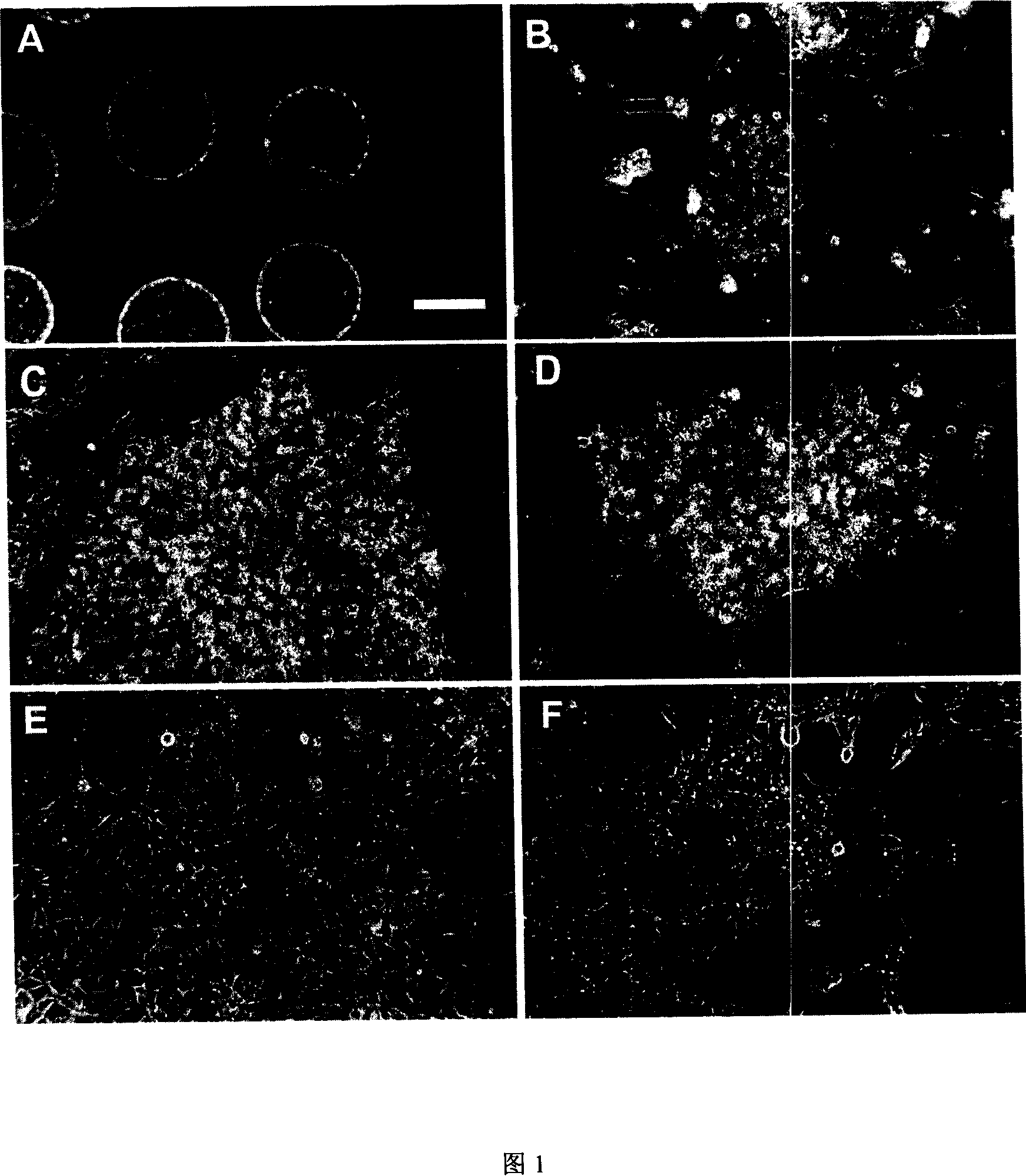
[0069] Example 1. rbES cell culture

[0070] In this example, we used basically the same isolation and culture methods to isolate embryonic stem cells from rabbit blastocysts of three subspecies (New Zealand, Angola, and Cinnamon). Among them, a cell line was obtained from New Zealand white rabbits, and One cell line was obtained from Angora rabbit, and three cell lines were obtained from Qingzilan rabbit (Figure 1 and Table 1). The successful establishment of rabbit embryonic stem cell lines from three different rabbit subspecies indicates that the method can be equally applied to other rabbit subspecies.

[0071] New Zealand, Angola, and Qingzilan rabbits were purchased from Shanghai Experimental Animal Center, Beijing Institute of Drug Control, and Nanjing Jinling Breeding Rabbit Farm. Four days after natural mating, the blastocysts were flushed out of the uterus with PBS, and the inner cell mass was isolated by mechanical or immunosurgical methods (Davor et al., 1975), a...

Embodiment 2

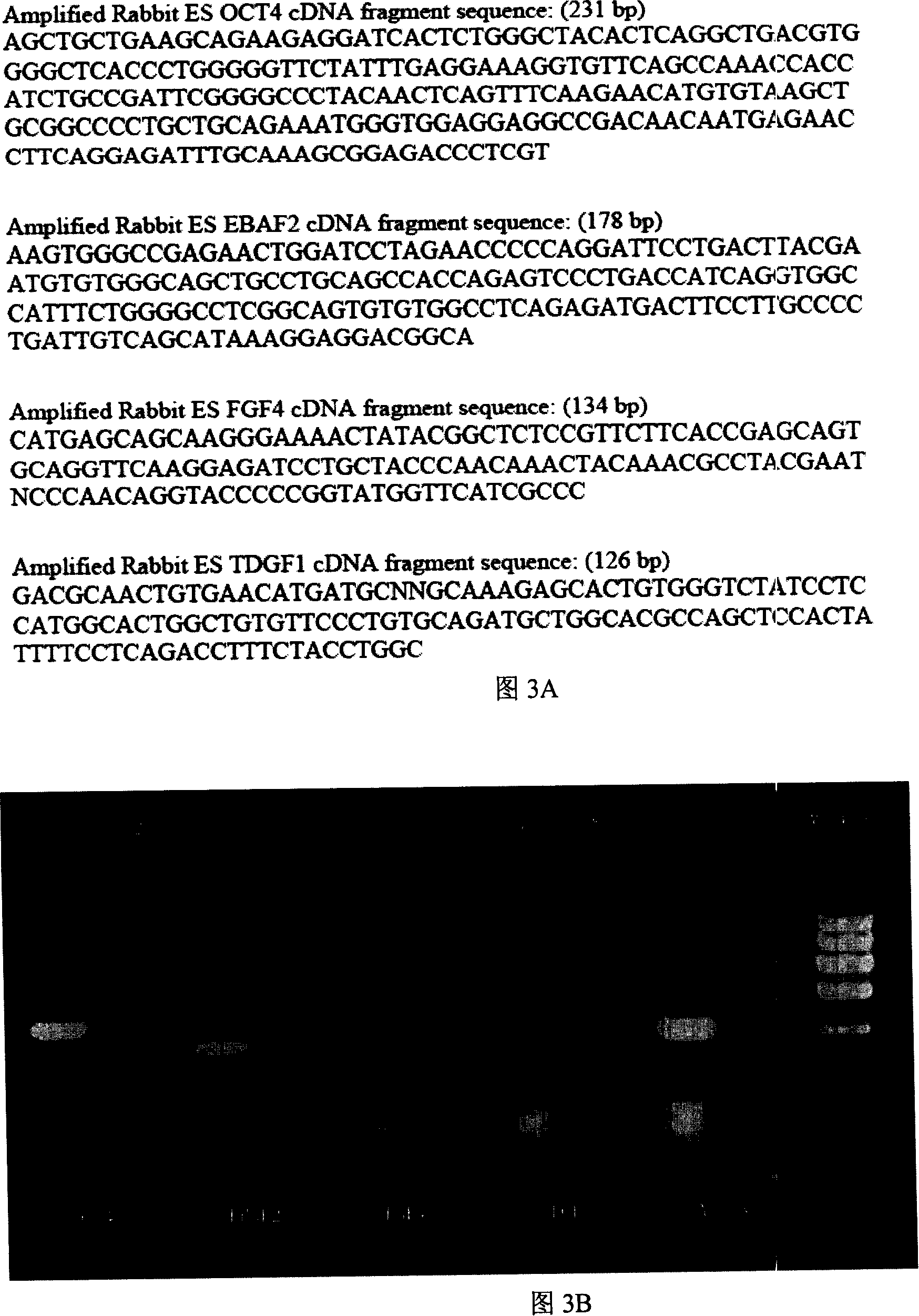
[0077] Example 2. Molecular markers expressed by rbES cells

[0078] RT-PCR

[0079] Total RNA was extracted with Trizol(R) (Invitrogen, Carlsbad, CA) and treated with DNase I at 37°C for 20 minutes to remove contaminating genomic DNA. First-strand synthesis was performed using Moloney murine leukemia virus reverse transcriptase (Promega, Madison, WI) and random primers (Promega) according to the protocol provided by the manufacturer. Primer sequences are shown in Table 4. PCR reactions were performed using the following parameters: denaturation at 94°C for 4 minutes, followed by 35 cycles of amplification consisting of denaturation at 94°C for 30 seconds, annealing for 30 seconds, extension at 72°C for 40 seconds, followed by a final extension at 72°C for 5 minutes. The annealing temperature is shown in Table 4. Samples without reverse transcriptase added during the reverse transcription step were used as controls for genomic DNA contamination. β-actin was used as an ...

Embodiment 3

[0088] Example 3. Calculation of cell cloning and population doubling time

[0089] We investigated the clonogenic rate of three rbES cell lines. Cell colonies were digested into single cells with 0.05% trypsin. 1000 cells per cell line were seeded into culture dishes. After 5 days, the number of colonies was counted, and the colony formation rate was calculated by the following formula: colony formation rate (%)=(clones / 1,000)×100%. As shown in Table 2, the colony formation rates of the three rbES cell lines rbES1, Qzl2 and LHR1 were 0.15±0.01%, 0.18±0.04% and 0.13+0.03%, respectively. The clonal growth of rbES cells will facilitate the selection of rbES cells in the process of genetic manipulation.

[0090] Table 2. Clonogenicity of rbES cells

[0091] cell line

Colony formation rate (%)

rbES1

0.15±0.01

Qz2

0.18±0.04

LHR1

0.13±0.03

[0092] Mark small (about 10 cells) cell colonies at the bottom of the culture...


PUM


Abstract

The present invention discloses a new method of obtaining rabbit embryonic stem cells (rbES), which adopts a serum free culture system to establish an rbES cell line which can proliferate under undifferentiated state for a long time (more than 1.5 years) and maintain the normal karyotype. The rbES expresses a pluripotent marker Oct4 and a plurality of other molecules, including EBAF2, FGF4, and TDGF1, which are expressed in stem cells. The stem cells can form an embryoid body in vitro and form a teratoma in immunodeficient mice. The rbES exhibits high colony forming efficiency and can be used as a nuclear donor to produce a clone rabbit, and genetic modifications of the rbES can be performed. The present invention also discloses potential applications of the rbES.

Description

Background of the invention [0001] Mice are widely used in biological and medical research due to several advantages. On the one hand, many wild-type congenic mouse strains with homologous genetic backgrounds have been established. On the other hand, many mouse models with genetic mutations, mostly obtained by homologous recombination techniques, have been established and characterized (Robertson et al., 1986; Thompson et al., 1989; Babinet et al., 2001) . Individual genes in these genetically mutant mice are deleted, increased, or modified, and such mice have a very important role in the field of genetic and developmental biology research. Some mice have genetic mutations that mimic human disease conditions (Holschneider et al., 2001; Harada et al., 1999; Kulkarni et al., 1999), and such mice provide valuable insights into the pathogenesis of human diseases. animal models. [0002] Although mice play an important role in genetic research, due to their small size, they are...

Claims


Application Information

Patent Timeline
IPC IPC(8): C12N5/06C12N5/0735
Inventor 方贞付李善刚陈学进盛慧珍
Owner XIN HUA HOSPITAL AFFILIATED TO SHANGHAI JIAO TONG UNIV SCHOOL OF MEDICINE
Why Eureka
Try Eureka