Processing method of CT cerebral hemorrhage image

A CT image and image technology, applied in the field of CT cerebral hemorrhage image processing, can solve problems such as the difficulty in establishing the spatial correspondence between normal brain atlas and abnormal brain tissue, brain tissue shift or deformation, and the impossibility of accurate realization.

Inactive Publication Date: 2008-08-13
SHENZHEN INST OF ADVANCED TECH
View PDF0 Cites 41 Cited by
  • Summary
  • Abstract
  • Description
  • Claims
  • Application Information

AI Technical Summary

Problems solved by technology

Methods that rely on more precise registration of brain atlas and data, where such registration is impossible to achieve precisely, for two reasons: First, due to the low spatial resolution of clinical CT data, it is not possible to precisely determine the Brain landmarks, second, brain tissue may shift or deform after hemorrhage, making it difficult to establish a spatial correspondence between normal brain atlas and abno

Method used


Image

  • Processing method of CT cerebral hemorrhage image
  • Processing method of CT cerebral hemorrhage image
  • Processing method of CT cerebral hemorrhage image

Examples


Embodiment Construction

[0061] Various preferred embodiments of the present invention will be described in detail below.

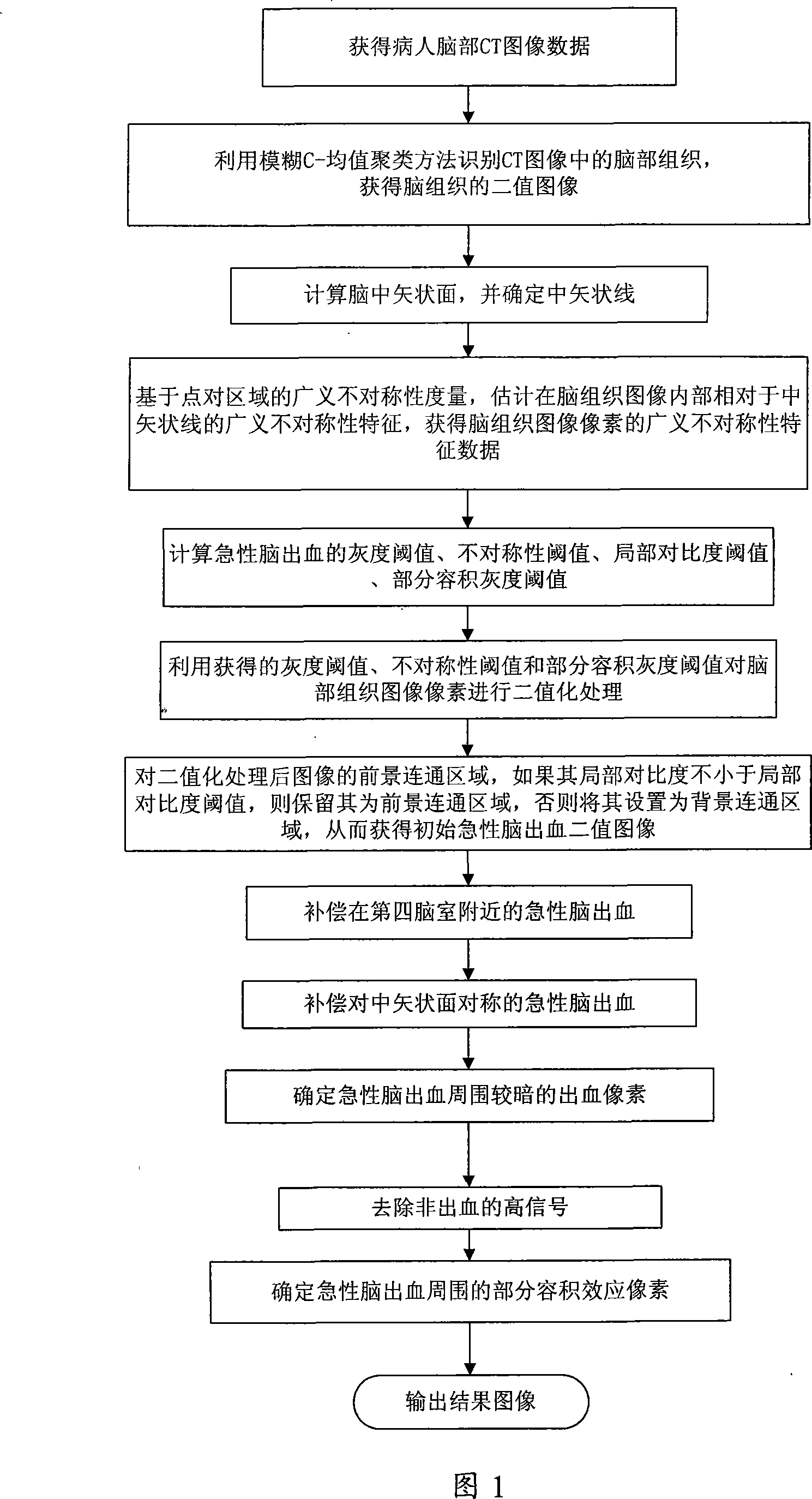
[0062] As shown in Figure 1, the processing method about CT cerebral hemorrhage image of the present invention can be used on the image processing system of general-purpose computer or CT image, carry out according to following step A to step G:

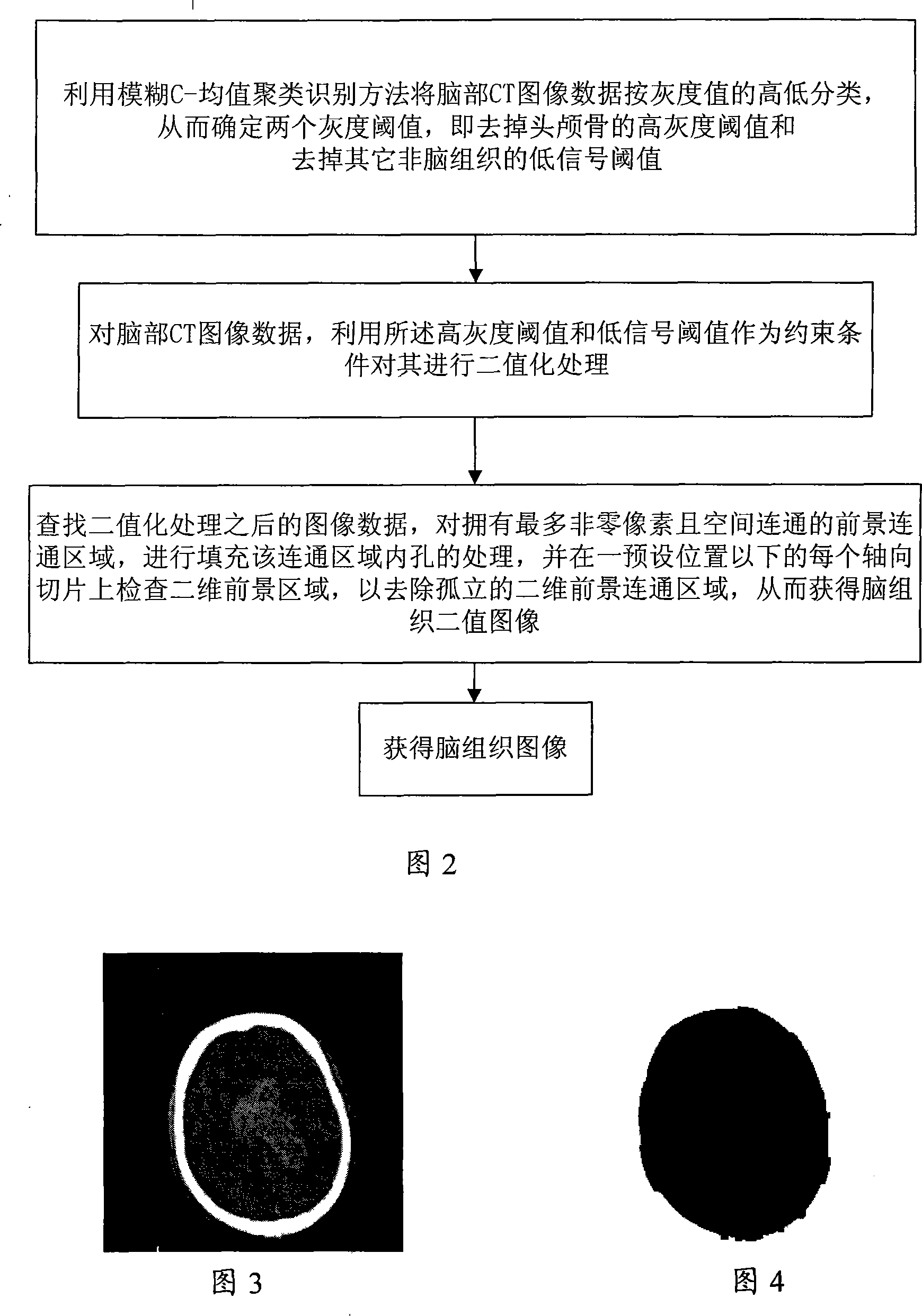
[0063] A. Use non-enhanced CT to obtain CT image data of the patient's brain. The CT data of the brain CT image (saved in standard DICOM format) is automatically converted into an 8-bit data file according to its window level and window width. Each image point Called a pixel (or voxel, hereinafter represented by a pixel), the grayscale of a pixel takes a value between 0 and 255. After obtaining multiple brain axial slices, sagittal slices or coronal slices, three-dimensional CT images can be obtained after conversion. The three-dimensional image coordinate system is: X is from left to right, Y is from front to back, and Z is from top to...


PUM


Abstract

The invention discloses a treating method of CT cerebral hemorrhage image, comprising: automatically selecting gray level image, binary image of brain tissue or binary image of head of raw data based on nonsingular number and winkling the brain median sagittal plane based on singular number; robustly depicting the asymmetry of instant cerebral hemorrhage to the median sagittal plane based on general asymmetry of dotted pairs area; adaptively figuring out gray threshold of instant cerebral hemorrhage, threshold of general asymmetry, local contrast threshold and partial bulk effect threshold; determining threshold of instant cerebral hemorrhage near the fourth ventricle by searching the axial slice below the pertrous bone; obtaining instant cerebral hemorrhage symmetrical to the median sagittal plane from the obtained unsymmetric hemorrhage; determining darker instant cerebral hemorrhage from determined lighter (higher gray scale) instant cerebral hemorrhage; removing high signal of not instant cerebral hemorrhage; determining instant cerebral hemorrhage pixel due to partial bulk effect, therefore the final instant cerebral hemorrhage image is obtained.

Description

technical field [0001] The invention relates to the technical field of CT image processing, in particular to a processing method for CT cerebral hemorrhage images, which is used to accurately determine the location of acute cerebral hemorrhage regions. Background technique [0002] Acute intracerebral hemorrhage refers to recent hemorrhage in the brain, which is a very urgent and dangerous sign, and its accurate and rapid detection is the prerequisite for effective treatment and treatment. Acute intracerebral hemorrhage is well identified by well-trained radiologists, but can also be erroneous when the hemorrhage is minimal or obscured by normal tissue; well-trained radiologists usually calibrate and calculate manually Cerebral hemorrhage volume, which is not only time-consuming, but also difficult to control accuracy, and the obtained scattered two-dimensional pictures are not the overall three-dimensional information. To make matters worse, when patients with bleeding ent...

Claims


Application Information

Patent Timeline
IPC IPC(8): A61B6/03G06T7/40
Inventor 胡庆茂
Owner SHENZHEN INST OF ADVANCED TECH
Why Eureka
Try Eureka