Progenitor endothelial cell capturing with drug eluting implantable medical device
A technology for medical equipment and drugs, applied in vascular endothelial cells, biochemical equipment and methods, drug delivery, etc., and can solve problems such as cracking
- Summary
- Abstract
- Description
- Claims
- Application Information
AI Technical Summary
Problems solved by technology
Method used
Image
Examples
Embodiment 1
[0172] The preparation of embodiment 1 coating composition
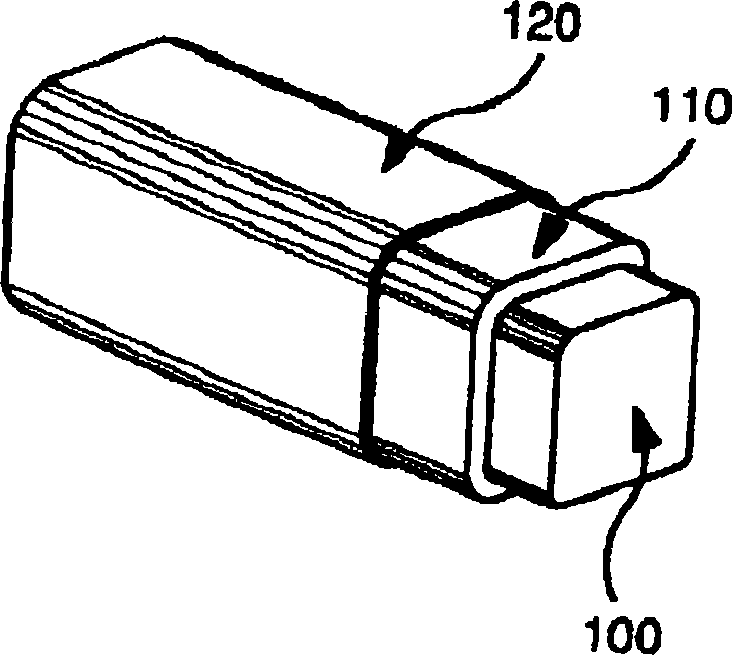
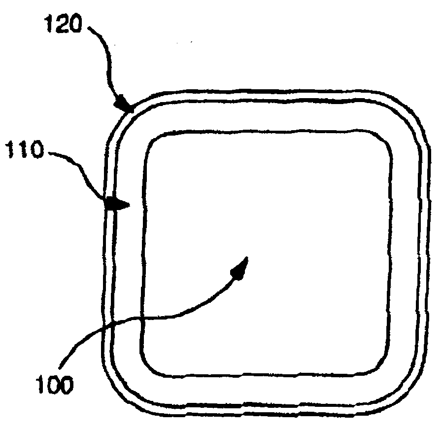
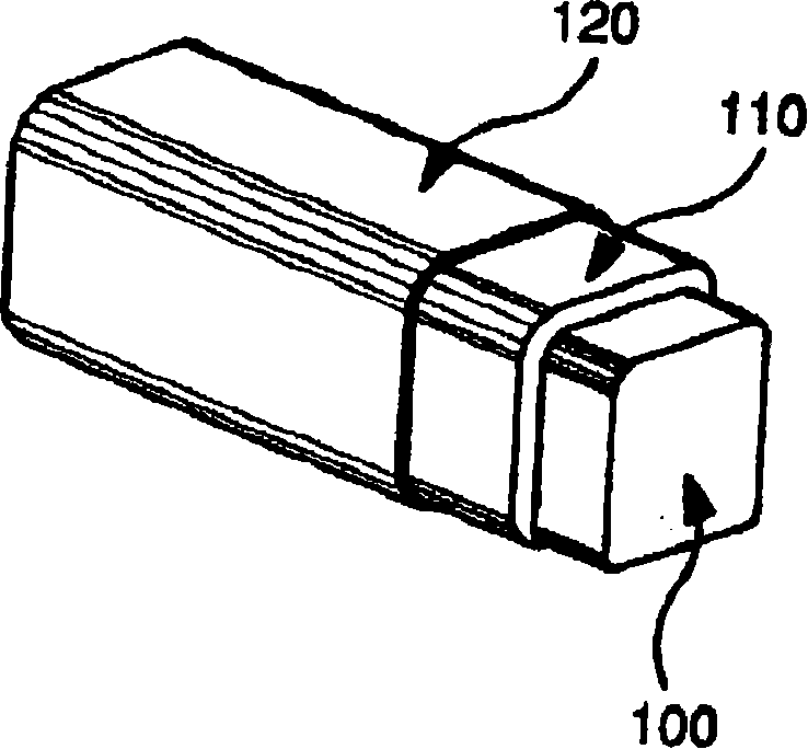
[0173] The polymer polyDL-lactide-co-glycolide (DLPLG, Birmingham Polymers) was provided as pellets. To prepare the polymer matrix composition for coating the stents, the pellets were weighed and dissolved in a ketone or dichloromethane solvent to form a solution. The drug was dissolved in the same solvent and added to the polymer solution to the desired concentration, thereby forming a uniform coating solution. The ratio of lactide to glycolide can be varied in order to improve ductility and alter the release kinetics of the coating matrix. This solution is then used to coat scaffolds to form Figure 11 Uniform coating as shown in . Figure 12 A cross-section through a coated stent of the invention is shown. The polymer / drug can be deposited on the surface of the stent using various standard methods.
Embodiment 2
[0174] Example 2 Evaluation of Polymers / Drugs and Concentrations
[0175] Method of spray-coating stents: polymer pellets of DLPLG that have been dissolved in a solvent are mixed with one or more drugs. Alternatively, one or more polymers can be dissolved with a solvent and one or more drugs can be added and mixed. The resulting mixture was evenly applied to the scaffold using standard methods. After coating and drying, the scaffolds were evaluated. The following list illustrates various examples of coating combinations that were investigated using various drugs and including DLPLG and / or combinations thereof. Alternatively, the formulation may consist of a base coat of DLPLG and a top coat of DLPLG or another polymer such as DLPLA and EVAC 25. Abbreviations for drugs and polymers used in coatings are as follows: MPA is mycophenolic acid, RA is retinoic acid; CSA is cyclosporine A; LOV is lovastatin.TM. (mevinolin)); PCT is paclitaxel; PBMA is polybutylmethacrylate, EVAC i...
Embodiment 3
[0200] The following experiments were performed to measure the drug elution profiles of coatings on stents coated by the method described in Example 2. The coating on the stent consisted of 4% paclitaxel and 96% of a 50:50 poly(DL-lactide-co-glycolide) polymer. Each scaffold was coated with 500ug of the coating composition and incubated in 3ml of bovine serum at 37°C for 21 days. Paclitaxel released into serum was measured on each day during the culture period using standard techniques. The experimental results are shown in Figure 13 middle. Such as Figure 13 As shown, the elution profile of paclitaxel release was very slow and controlled as only about 4ug of paclitaxel was released from the stent over the 21 day period.
PUM
| Property | Measurement | Unit |
|---|---|---|
| pore size | aaaaa | aaaaa |
| pore size | aaaaa | aaaaa |
| thickness | aaaaa | aaaaa |
Abstract
Description
Claims
Application Information
 - R&D
- Intellectual Property
- Life Sciences
- Materials
- Tech Scout
- Unparalleled Data Quality
- Higher Quality Content
- 60% Fewer Hallucinations
Browse by: Latest US Patents, China's latest patents, Technical Efficacy Thesaurus, Application Domain, Technology Topic, Popular Technical Reports.
© 2025 PatSnap. All rights reserved.Legal|Privacy policy|Modern Slavery Act Transparency Statement|Sitemap|About US| Contact US: help@patsnap.com


