Polar sensitive fluorescent probe compounds for arginine residue marker, preparation and use thereof

An arginine and residue technology, applied in the field of new polarity-sensitive fluorescent probe compounds, can solve the problem of no fluorescence and achieve the effect of stable fluorescence properties

Inactive Publication Date: 2008-12-17
INST OF CHEM CHINESE ACAD OF SCI
View PDF0 Cites 2 Cited by
  • Summary
  • Abstract
  • Description
  • Claims
  • Application Information

AI Technical Summary

Problems solved by technology

However, these reagents are not inherently fluorescent, so they cannot be detected using fluorescent methods

Method used


Image

  • Polar sensitive fluorescent probe compounds for arginine residue marker, preparation and use thereof
  • Polar sensitive fluorescent probe compounds for arginine residue marker, preparation and use thereof
  • Polar sensitive fluorescent probe compounds for arginine residue marker, preparation and use thereof

Examples


Embodiment 1

[0021] Example 1: 3-(4-chloro-6-p-formylformylphenoxy-1,3,5-triazineamino)-7-dimethylamino-2-methylphenazine (CGTDP) preparation

[0022] 3-(4-chloro-6-p-formylformylphenoxy-1,3,5-triazine amino)-7-dimethylamino-2-methylphenazine of the present invention consists of three parts: Radix Red is used as a polar sensitive group, cyanuric chloride is used as a bridging agent, and phenylglyoxal is used as an arginine labeling group. The synthetic route is as follows:

[0023]

[0024] The specific synthesis process is as follows:

[0025] Step a): Dissolve 280 mg (1.5 mmol) of cyanuric chloride in about 10 ml of ice acetone, slowly add about 40 ml of neutral red (288 mg, 1 mmol) in ice acetone and 4 ml of sodium carbonate (106 mg, 1 mmol) in water, The pH of the reaction solution is between 8 and 9, the time for dropping is about 20 minutes, and the reaction is stirred at 0 to 5°C. After reacting for 1.5 h, ice was added to stop the reaction, and the resulting precipitated prod...

Embodiment 2

[0028] Spectral properties of embodiment 2, CGTDP

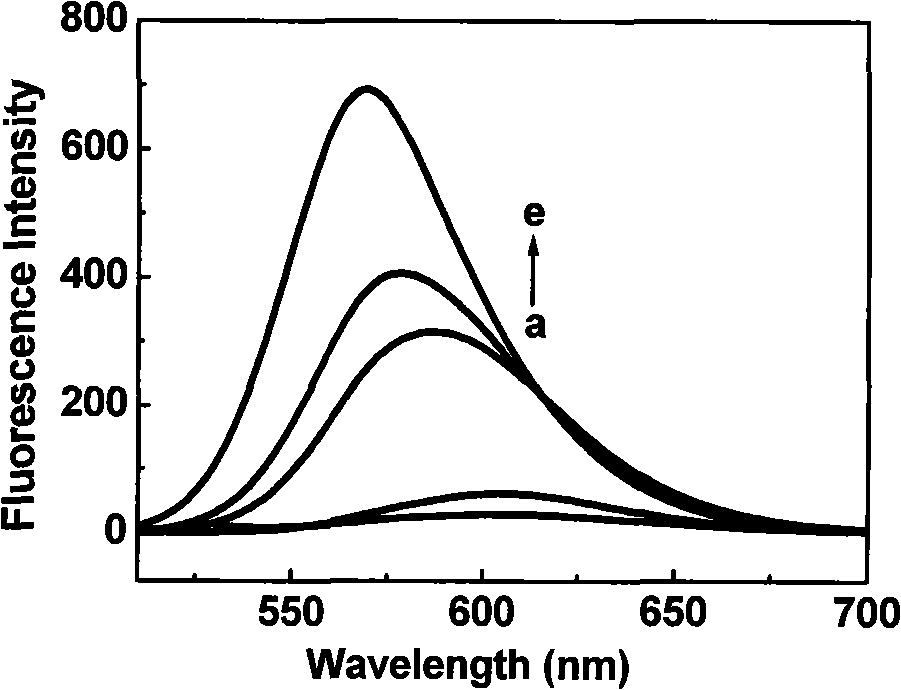
[0029] Dissolve CGTDP in different solvents (concentration is 10 μ M), measure its fluorescence emission spectrum, the result is as follows figure 1 shown. When measuring the fluorescence emission spectrum, the corresponding maximum excitation wavelength was used to excite; the slit width of excitation and emission was 10nm. In the figure, (a) water (0.1M NaHCO 3 , pH 8.6); (b) dimethylsulfoxide; (c) acetonitrile; (d) acetone; (e) tetrahydrofuran.

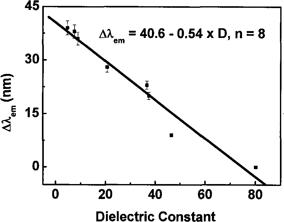
[0030] The spectral data of CGTDP (10 μ M) in different solvents are shown in Table 1, and its maximum emission wavelength shift value (Δλ em ) and solvent dielectric constant (D) linear relationship diagram as shown figure 2 As shown, the linear equation between the fluorescence maximum emission wavelength shift value and the dielectric constant: Δλ em =40.6-0.54×D.

[0031] Table 1. Fluorescence spectral properties of CGTDP in different solvents

[0032]

[0033] im...

Embodiment 3

[0036] Example 3: Using the polarity-sensitive fluorescent probe CGTDP to selectively label the arginine residue in the active site of creatine kinase (CK) and detect the polarity near the active site.

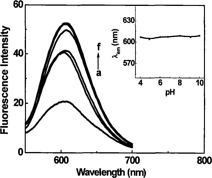
[0037] Prepare 2 ml of creatine kinase CK (5 μM) in sodium bicarbonate buffer solution (pH 8.6), add 100 μl of CGTDP (4 mM) in DMSO solution, and react at 30° C. for 1.5 h. The protein-labeled products and free fluorescent reagents in the reaction solution were separated by Sephadex G-25 gel column, and detected by UV-visible spectrophotometer, and the labeled protein part with simultaneous absorption at 280nm and 508nm was collected.

[0038] Figure 5 is the UV absorption spectrum of CGTDP, creatine kinase, and creatine kinase labeled with CGTDP. In the figure, (a) CGTDP (2.75 μM), (b) creatine kinase (2.2 μM), (c) creatine kinase labeled with CGTDP Acid kinase (2.3 μM). It can be seen from the figure that the simultaneous appearance of absorption peaks around 280nm and 50...


PUM


Abstract

The invention discloses a polarity-sensitive fluorescent probe compound applicable to marking arginine residue and a preparation method and application thereof. The fluorescent probe is 3-(4-chlorin-6-P-Toluoyl formylphenyl phenoxy-1, 3, 5-triazine amino)-7-dimethylamino-2-toluphenazine and the structure thereof is as formula I. The invention prepares a polarity-sensitive fluorescent probe which can be used for selectively marking compounds containing the arginine residue; owing to introduction of phenylglyoxal group, the fluorescent probe can be used for marking biomacromolecule containing the arginine residue and determining and researching the changing situation of the polarity in the area near the biomacromolecule, in particular to selectively marking the arginine residue in protein and researching the changing situation of the polarity in the area near the arginine residue.

Description

technical field [0001] The present invention relates to a novel polarity-sensitive fluorescent probe compound suitable for arginine residue labeling——3-(4-chloro-6-p-formylformylphenoxy-1,3,5-tri Azinamino)-7-dimethylamino-2-methylphenazine, and preparation method and application thereof, particularly relate to the use of the compound in labeling arginine residues in proteins and detecting the extremes near the active sites of many enzymes sex application. Background technique [0002] The structure and function of protein molecules are closely related, and its local misfolding can cause diseases related to it. Therefore, the development of regional structure analysis and research methods of protein molecules (such as acquisition of local information, research on folding or local conformational changes, etc.) will help to understand the essence of life and the mechanism of disease occurrence at the molecular level. In the study of the structure and function of enzymes, the...

Claims


Application Information

Patent Timeline
IPC IPC(8): C07D403/12G01N33/52
Inventor 马会民王淑娟
Owner INST OF CHEM CHINESE ACAD OF SCI
Why Eureka
Try Eureka