Bionic dermis as well as preparation method and application thereof
A dermal and bionic technology, applied in the field of medical and surgical repair materials, can solve the problems of lack of mechanical properties of skin and dermis
- Summary
- Abstract
- Description
- Claims
- Application Information
AI Technical Summary
Problems solved by technology
Method used
Image
Examples
Embodiment 1
[0077] Preparation of Bionic Dermis I
[0078] Normal tissue skin was decellularized and then homogenized. This normal tissue contains various cell growth factors mentioned above, proteoglycans, glycoproteins, and protein molecules that promote cell and fiber adhesion.
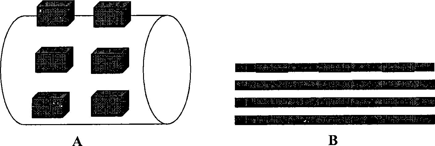
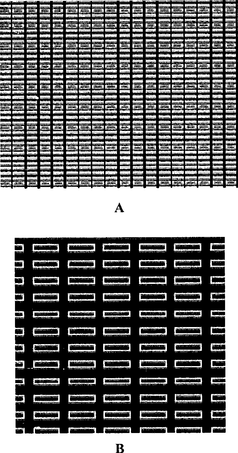
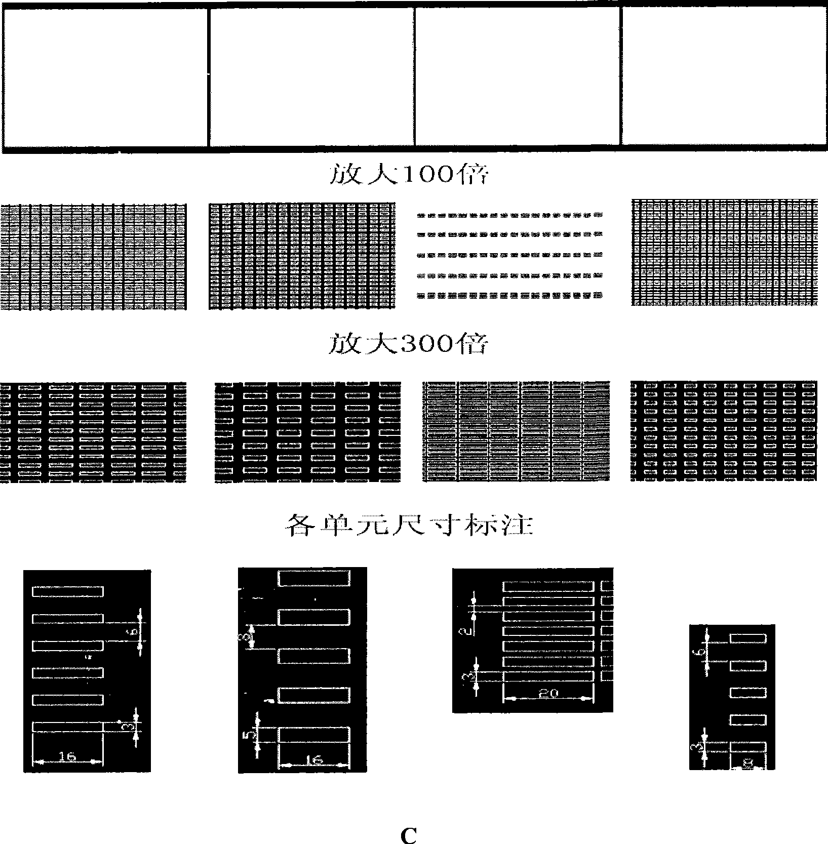
[0079] Three-dimensional multi-directional (4, 5, 6, 7) weaving of different fibers, and then immersed in the homogenate prepared above, at room temperature, after 30 minutes, taken out and dried. Get the dermis template as described. The area can be determined according to the area of the wound.
[0080] Then the dermal template was implanted into the wound, the superficial blade-thick skin was grafted, and bandaged under pressure.
Embodiment 2
[0082] Preparation of Bionic Dermis II
[0083] Dip different fibers 3, 4, 5, 6, 7, 8, 9, 10, and 11 into fibronectin (20-200 micrograms / ml), collagen (10-100 micrograms / ml), in vitro fibronectin (vitronectin, VN) (1-50 micrograms / ml), hyaluronic acid (2-50 micrograms / microliter), non-collagenous glycoproteins, elastin, syndecan, aminoglycans, glycoproteins and growth factors, and Immerse in PEG or PLL-g-PEG (1mg / ml), then take it out, dry it, make different combinations, three, or five or seven, each combination contains PEG or PLL-g-PEG, and then edit The braids are made into fiber bundles, and then the fiber bundles are three-dimensionally multi-directional, five-directional, and seven-directional to weave to make a leather template (that is, bionic leather II). The area can be determined according to the area of the wound.
[0084] Then the dermal template was implanted into the wound, the surface was grafted with autologous slash-thick skin, and bandaged under pressur...
Embodiment 3
[0088] Animal experiments of bionic leather
[0089] In this embodiment, animal experiments are used to test, and the test method is as follows: select a certain number of clean Duroc pigs (purchased from: Shanghai Slack Experimental Animal Co., Ltd.), 24 hours after the back hair removal, use a scalpel on the back of the pig A full-thickness skin of a certain size was cut, and the blade-thick skin was taken back on a drum-type dermatome as autologous skin. The wounds were randomly divided into four groups:
[0090] The wound in group 1 is the experimental wound. After hemostasis, the bionic dermis prepared in Example 1 was first transplanted, and then the autologous skin was transplanted on it, and bandaged under pressure;
[0091] The wound in group 2 is the experimental wound. After hemostasis, the bionic dermis prepared in Example 2 was first transplanted, and then the autologous skin was transplanted on it, and bandaged under pressure;
[0092] The wound in group 3 was ...
PUM
 Abstract
Description
Claims
Application Information
 - R&D
- Intellectual Property
- Life Sciences
- Materials
- Tech Scout
- Unparalleled Data Quality
- Higher Quality Content
- 60% Fewer Hallucinations
Browse by: Latest US Patents, China's latest patents, Technical Efficacy Thesaurus, Application Domain, Technology Topic, Popular Technical Reports.
© 2025 PatSnap. All rights reserved.Legal|Privacy policy|Modern Slavery Act Transparency Statement|Sitemap|About US| Contact US: help@patsnap.com


