Method for producing brain sample of small animal

A technology of small animals and specimens, which is applied in the field of preparation of whole brain specimens of small animals, can solve the problems of being unable to observe the three-dimensional shape of whole brain neurons and the complete shape of whole brain neurons, and achieve the effect of high contrast and not easy to fade

Inactive Publication Date: 2009-06-17
HUAZHONG UNIV OF SCI & TECH
View PDF0 Cites 14 Cited by
  • Summary
  • Abstract
  • Description
  • Claims
  • Application Information

AI Technical Summary

Problems solved by technology

[0014] The above methods can meet many needs, but have the following disadvantages: it is impossible to observe the three-dimensional morphology of whole brain neurons in three directions with the same submicron resolution
Although submicron sections can be obtained from resin section specimens, the specimens produced are not whole brain specimens, and the staining method only stains neuron cell bodies but not neuron dendrites and axons, so it is impossible to observe the complete morphology of whole brain neurons

Method used


Image

  • Method for producing brain sample of small animal
  • Method for producing brain sample of small animal

Examples


Embodiment 1

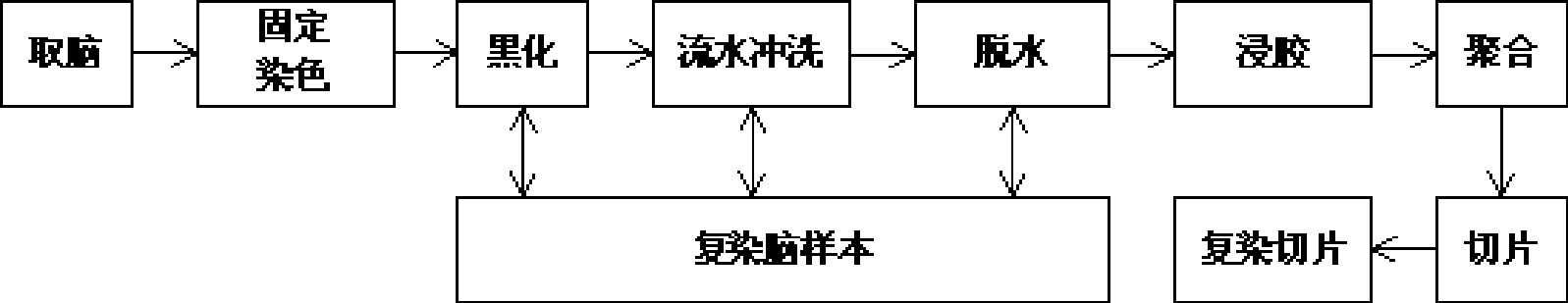
[0042] 1. Material collection: Adult mice were taken, decapitated after deep anesthesia, and the whole brain was dissected out;

[0043] 2. Fixation and staining: Fresh whole brains were immediately put into the fixative solution for fixation and staining. The dipping temperature was 0°C. After staining for 90 days, the solution was replaced regularly. The solution was changed once after fixing for 1 day, and the solution was changed every 20 days thereafter, at least The medium was changed 3 times, and the formula of the fixative solution was:

[0044] Mercury: 7% by weight

[0045] Potassium dichromate: 7% by weight

[0046] Potassium chromate: 5% by weight

[0047] Distilled water: 81% by weight

[0048] The fixative solution must be prepared now, and potassium chromate solution must be added at the end. The fixative solution must be packed in a brown bottle, wrapped in tin foil and kept in a dark place to reduce the influence of light, and the reagent bottle should be s...

Embodiment 2

[0059] 1. Material collection: Adult mice were taken, decapitated after deep anesthesia, and the whole brain was dissected out;

[0060] 2. Fixation and staining: Fresh whole brains were immediately put into the fixative solution for fixation and staining. The dipping temperature was 25°C. After staining for 120 days, the solution was replaced regularly. The medium was changed 3 times, and the formula of the fixative solution was:

[0061] Mercury: 10% by weight

[0062] Potassium dichromate: 10% by weight

[0063] Potassium chromate: 10% by weight

[0064] Sodium tungstate: 5% by weight

[0065] Distilled water: 65% by weight

[0066] The fixative solution must be prepared now, and potassium chromate solution must be added at the end. The fixative solution must be packed in a brown bottle, wrapped in tin foil and kept in a dark place to reduce the influence of light, and the reagent bottle should be shaken frequently;

[0067] 3. Blackening: soak the stained whole brain ...

Embodiment 3

[0079] 1. Material collection: Frogs were taken, decapitated after deep anesthesia, and the whole brain was dissected out;

[0080] 2. Fixation and staining: Fresh whole brains were immediately put into the fixative solution for fixation and staining. The dipping temperature was 50°C. Dyeing was carried out for 120 days, and the solution was replaced regularly. The medium was changed 3 times, and the formula of the fixative solution was:

[0081] Mercury: 0.1% by weight

[0082] Potassium dichromate: 1.5% by weight

[0083] Potassium chromate: 1% by weight

[0084] Potassium tungstate: 0.1% by weight

[0085] Distilled water: 97.3% by weight

[0086] The fixative solution must be prepared now, and potassium chromate solution must be added at the end. The fixative solution must be packed in a brown bottle, wrapped in tin foil and kept in a dark place to reduce the influence of light, and the reagent bottle should be shaken frequently;

[0087] 3. Blackening: soak the stain...


PUM


Abstract

The invention discloses a method for preparing a whole brain specimen of a baby animal. The steps are as follows: a. a baby animal brain sample is fixed and imbued; b. the fixed and imbued brain sample is blackened by using blackening liquor that is alkali liquor with the pH value being more than 10, and the blackening period is 1-24 hours; c. the obtained brain sample is dehydrated, and the dehydration is carried out by 100% dehydrating agent in weight; d. the blackened brain sample is gummed by using an embedding agent, and two continuous gumming are carried out by 100% embedding agent in weight; e. after being gummed by the embedding agent, the brain sample is put into an embedding box and then submerged by adding the 100% embedding agent in weight; f. the embedding box for submerging the brain sample is heated, so that the embedding agent is polymerized, the polymerization temperature is 35 to 80 DEG C, and the polymerization period is 6-96 hours. The method for preparing the baby animal brain specimen of the invention can dye more neurons as well as neuron cell bodies, dendrite and axon; the contrast is high; the color is not faded easily; and whole brain neuron three dimensional morphology structures can be observed in the sub-micro scale.

Description

technical field [0001] The invention relates to a preparation method of a specimen, in particular to a preparation method of a small animal whole brain specimen. Background technique [0002] It is widely used in clinical diagnosis and scientific research to make stained samples of brain neurons and observe neuron morphology. It mainly involves two techniques: neuronal staining and specimen embedding techniques. Among them, neuron staining techniques mainly include two types: Golgi-Cox staining and common dyes (such as hematoxylin, methylene blue) staining. Embedding techniques are mainly divided into paraffin embedding, collodion embedding and resin embedding according to different embedding agents. [0003] According to different combinations of staining techniques and embedding techniques, the commonly used neuron staining specimen preparation methods mainly include the following categories: [0004] Paraffin section specimens: Paraffin specimens are usually prepared f...

Claims


Application Information

Patent Timeline
Patent Type & Authority Applications(China)
IPC IPC(8): G01N1/30G09B23/28
Inventor 骆清铭张斌李安安龚辉王冰然
Owner HUAZHONG UNIV OF SCI & TECH
Why Eureka
Try Eureka