Protein suspending chip for composite detection of multiple kinds of pathogens, its production method and detection method

A suspension chip and composite detection technology, which is applied in the direction of measuring devices, biological testing, material inspection products, etc., can solve the problems of lack of models and evaluations, and achieve the effects of simple preparation methods, wide dynamic range, and good applicability

Inactive Publication Date: 2009-08-05
CHINESE ACAD OF INSPECTION & QUARANTINE
0 Cites 8 Cited by

AI-Extracted Technical Summary

Problems solved by technology

However, whether the suspension chip can detect multiple types of pathogens at the same time, whether it is suitable for rapid and direct detectio...

Abstract

The invention relates to a method for preparing and detecting a protein suspension chip for compound detection of various pathogens. The pathogens comprise Yersinia pestis, Bacillus anthracis, staphylococcal enterotoxin B, SEB, ricin and SARS-CoV. Substantive tests prove that the method for preparing the protein suspension chip is simple, convenient and rapid and has rapid and accurate detection effect, high sensitivity, wide range of detection, strong specificity and favorable applicability for actual environment samples. And meanwhile, the invention lays a foundation for setting up a multifunctional, multi-index, high-flux and standardized requirement for the compound detection of the pathogens.

Application Domain

Chemiluminescene/bioluminescenceBiological testing

Technology Topic

PathogenProtein formation +7

Image

  • Protein suspending chip for composite detection of multiple kinds of pathogens, its production method and detection method
  • Protein suspending chip for composite detection of multiple kinds of pathogens, its production method and detection method
  • Protein suspending chip for composite detection of multiple kinds of pathogens, its production method and detection method

Examples

  • Experimental program(5)

Example Embodiment

[0044] 2. Preparation of samples to be tested
[0045] 1. Preparation of samples for single component analysis
[0046] The concentration of Y. pestis stock solution is 10 8 cfu/mL, the concentration of anthrax spore stock solution is 10 7 cfu/mL, the stock solution concentration of ricin, SEB, and SARS-CoV N protein is 1mg/mL. The toxin and protein samples are diluted before use. The concentration range of the bacterial suspension is 10 1 -10 8 cfu/mL, the spore concentration range is 10 2 -10 7 cfu/mL, the concentration range of toxin and protein is 10pg/mL-5μg/mL. The bacteria to be analyzed are diluted with PB into 10-fold different gradients, and toxins and proteins are diluted with PB into 4-fold different gradients. The concentration of several samples is lower than the detection sensitivity. High-concentration samples should make the binding site of the coded microspheres in Saturated state.
[0047] 2. Preparation of mixed samples
[0048] The mixed sample contains two to five of anthracis spore, Yersinia pestis, SARS-CoV N protein, ricin and SEB, which are respectively diluted from the corresponding stock solution with sample diluent and mixed to prepare. Mixed samples include samples for multiple tests of pathogens, with different components and ratios, and random combinations.
[0049] 3. Preparation of simulated pollution samples
[0050] Add 0.5g of milk powder, corn starch, wheat flour, instant fruit powder and other powders to 5mL sample diluent (PB buffer), and add different concentrations of anthrax spore, Yersinia pestis, SARS-CoVN protein, ricin and SEB One or more of them are mixed into the powder sample, thoroughly shaken and mixed, and allowed to stand for more than 2 hours to fully adsorb the target analyte and the simulated white powder. After filtering with absorbent cotton, thin filter paper, thick filter paper, 0.45μm filter membrane filter paper or low speed centrifugation (1000rpm, 1min), the supernatant is used as the sample to be tested for the detection of the suspension chip method.
[0051] 4. Preparation of blind samples
[0052] A total of 46 samples of single analyte samples, mixed samples and simulated contamination samples from different media were drawn and prepared, and the sequence and numbering were disturbed, and the detection was performed as a blind sample. Blind samples include 8 blank or other interference samples, 38 samples containing test substances, including 11 single-factor analytes in the sample treatment solution, 13 mixed samples, and 14 simulated pollution samples (including 5 mixed samples).

Example Embodiment

[0053] Example 1. Preparation of protein suspension chip
[0054] A. Activation of coded microspheres
[0055] Select 5 kinds of microspheres to label Y. pestis antibody (No. 028), anthrax spore antibody (No. 025), SEB antibody (043), ricin antibody (027), SARS-CoV N protein antibody (No. 044), take 100 μL (1.25×10 6 A) The encoded microspheres are placed in a 1.5mL centrifuge tube, centrifuged at 14000g, carefully aspirate and discard the supernatant. Add 100 μL of microsphere washing buffer to suspend, shake and sonicate, centrifuge at 14000g, carefully aspirate and discard the supernatant. Add 100μL of microsphere activation buffer, then add 10μL of freshly prepared EDC (50mg/mL), and then add 10μL of freshly prepared 50mg/mL carboxyl-active biotin (ie Sulfo-NHS-Biotin, SH-active Biotin), shake for 20 minutes at room temperature. Add 150 μL of PBS (pH 7.4), shake, centrifuge at 14000g, aspirate carefully and discard the supernatant. Add 100 μL of PBS (pH 7.4) to suspend the coded microspheres.
[0056] B. Antibody-coated coded microspheres
[0057] Separately take 10 μg of each target detection substance capture antibody (as shown in Table 1) and add them to the activated coded microspheres, dilute to 500 μL with PBS buffer, and shake at room temperature for 2 hours. Centrifuge at 14000g, aspirate carefully and discard the supernatant. Wash once with 500μL of PBS buffer, centrifuge at 14000g, aspirate carefully and discard the supernatant. Add 250 μL of blocking buffer to suspend the coded microspheres, shake at room temperature for 30 minutes, centrifuge at 14000g, aspirate carefully and discard the supernatant. Add 500 μL of microsphere storage solution to wash the coded microspheres, centrifuge at 16000g, carefully aspirate and discard the supernatant. Finally, the coded microspheres were suspended in 150μL of microsphere preservation solution and stored at 4°C in the dark for later use.
[0058] C. Counting of coated microspheres
[0059] Take appropriate amount of microspheres and dilute them with a hemocytometer (0.10mm; 1/400mm 2) Count under an ordinary microscope. According to the formula (each large grid number × 10 4 × Dilution times × volume (mL)) to calculate the number of microspheres.
[0060] D. Biotinylation of detection antibodies
[0061] Prepare a concentration of 10mM biotin solution and 2mg/mL of each target detection antibody solution to be labeled (as shown in Table 1), add the calculated volume of biotin to the antibody solution to be labeled, and shake at room temperature for 30 minutes (Or on ice for 2 hours), pass the column to desalinate and then aliquot, and store at -20℃ for later use.
[0062] The calculation process of antibody dosage is as follows:
[0063] Take 1mL solution of IgG (molecular weight 150,000) labeled with 2mg/mL as an example, it is necessary to add about 27μ1 of 10mM biotin solution.
[0064] 1 ml lgG × 2 mg lgG 1 ml lgG × 1 m mol lgG 150,000 mg lgG × 20 mmol Biotin 1 m mol lgG = 0.000266 m mol Biotin
[0065] 0.000266 mmol Biotin × 1,000,000 μl L × L 10 mmol = 26.6 μlBiotin Reagent
[0066] Among them, biotin is biotin.

Example Embodiment

[0067] Example 2. Improvement of sensitivity and dynamic detection range of target pathogens
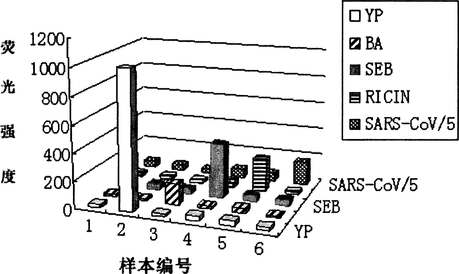
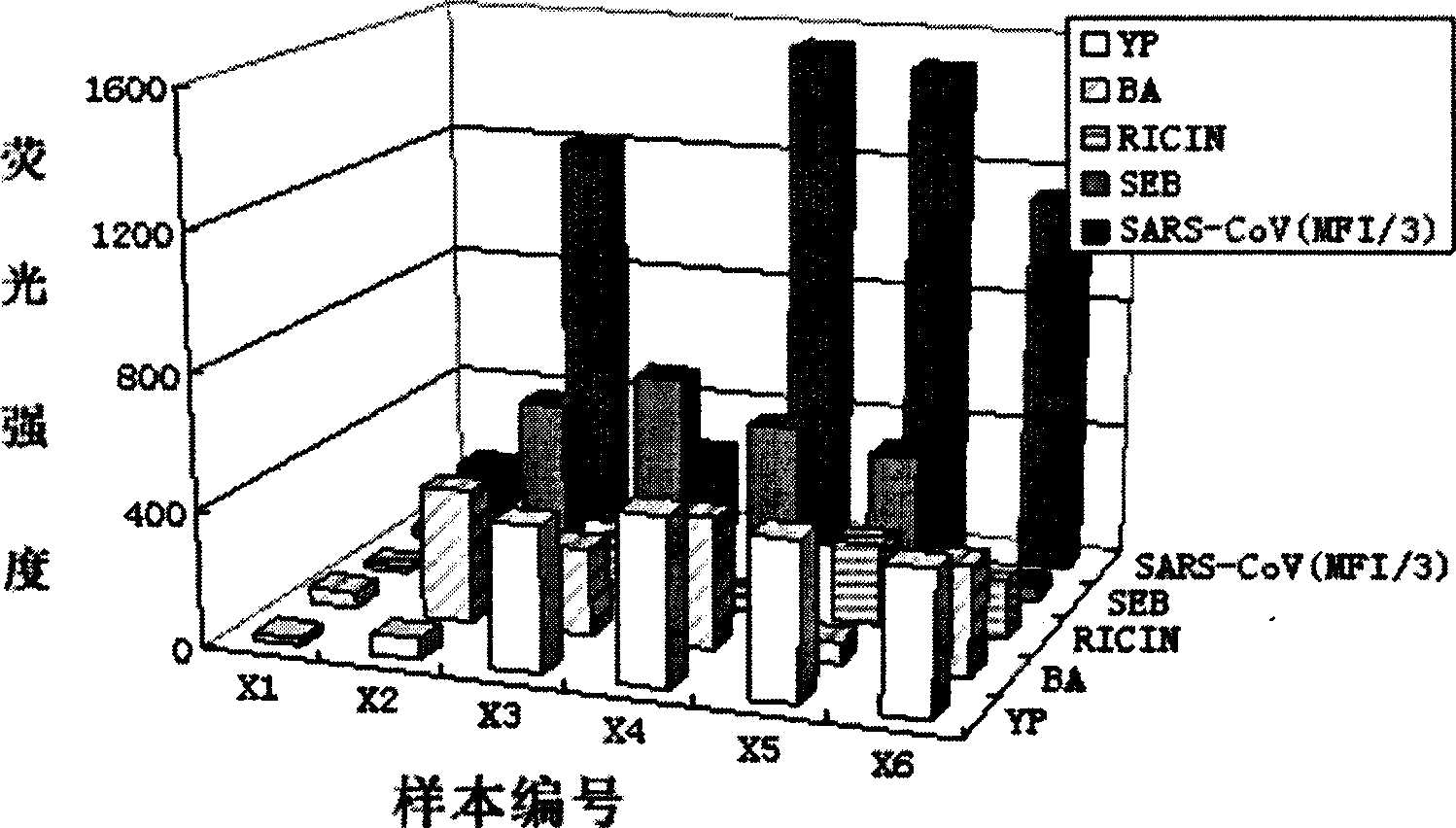
[0068] A. Preparation of samples to be tested
[0069] Separate the Y. pestis stock solution (10 8 cfu/mL) and anthrax spore stock solution (10 7 cfu/mL) was diluted with PB 10-fold to form a series of concentration gradient samples, and ricin, SEB, SARS-CoVN protein (stock solution 1mg/mL) was diluted with PB 4-fold to form a series of concentration gradient samples. Make the Yersinia pestis suspension in the range of 10 1 ~10 8 cfu/mL, the concentration range of anthrax spores is 10 2 ~10 7 The concentration range of cfu/mL, ricin, SEB, and SARS-CoV N protein is 10pg/mL~5μg/mL.
[0070] B. Testing of samples
[0071] All reactions in the detection process are carried out on 96-well filter plates, and the detection process is as follows:
[0072] (1) Add 50μL of working solution containing corresponding coded microspheres to each well, wash with cleaning solution and filter with vacuum pump;
[0073] (2) Add 50μL of test sample, mix well and shake for 30 minutes at room temperature, avoid light, wash with cleaning solution and filter with suction;
[0074] (3) Add 50 μL of the biotinylated antibody diluted with the antibody diluent at an appropriate concentration, mix and shake at room temperature for 30 minutes in the dark, wash the lotion and filter by vacuum pump;
[0075] (4) Add 50μL of SA-PE, mix well and shake at room temperature for 10 minutes. Wash the lotion and filter by vacuum pump;
[0076] (5) Add 125μL of detection buffer, resuspend and mix by shaking;
[0077] (6) Use the floating chip system to read the FMI value and analyze the data.
[0078] 3. Determination of the detection sensitivity and detection range of the target pathogen
[0079] The lowest detection limit (LOD value) of the protein suspension chip detection method is the concentration of the detection substance corresponding to the cutoff value of the detection fluorescence intensity. Among them, the definition of Cutoff is to use the blank control sample (Blank) fluorescence detection signal MFI mean plus 3 times the standard deviation (SD), that is, Cutoff value = MFI Blank +3×SD. If the detection result is higher than the fluorescence intensity value corresponding to the LOD, it is determined that the target detection result is positive; if the detection result is lower than the fluorescence intensity value corresponding to the LOD, it is determined that the target detection result is negative. The highest detection limit is the concentration of the test substance that makes the binding site of the coded microsphere in a saturated state, that is, the MFI value obtained from the detection begins to enter a plateau with the increase of the sample concentration, indicating that the concentration of the test substance in the sample is too high, and the sample needs to be diluted Check again later. Therefore, according to the minimum detection limit and the maximum detection limit, the sensitivity and dynamic detection range of the protein suspension chip to detect the target pathogen can be determined. The detection results are shown in Table 3.
[0080] Table 3 Sensitivity of protein suspension chip method to detect five pathogens
[0081]
[0082] In conclusion, the detection sensitivity and detection range of the protein suspension chip of the present invention for the above five pathogens are significantly improved compared to the ELISA method.

PUM

no PUM

Description & Claims & Application Information

We can also present the details of the Description, Claims and Application information to help users get a comprehensive understanding of the technical details of the patent, such as background art, summary of invention, brief description of drawings, description of embodiments, and other original content. On the other hand, users can also determine the specific scope of protection of the technology through the list of claims; as well as understand the changes in the life cycle of the technology with the presentation of the patent timeline. Login to view more.

Similar technology patents

Method for preparing catalyst with non-uniform distribution of active metal component

InactiveCN101462080AEasy to prepareHigh hydrodemetallization activity
Owner:CHINA PETROLEUM & CHEM CORP +1

Medical adhesives and preparation method thereof

ActiveCN105327388AEasy to prepareshort gel time
Owner:莫秀梅 +1

Classification and recommendation of technical efficacy words

  • high sensitivity
  • Easy to prepare

System and method for optical coherence imaging

ActiveUS20060055936A1sufficient signal to noise ratiohigh sensitivity
Owner:THE GENERAL HOSPITAL CORP

Three-axis integrated MEMS accelerometer

InactiveUS20060272413A1low costhigh sensitivity
Owner:VAGANOV VLADIMIR +1

Capacitive micromachined acoustic transducer

ActiveUS20060233401A1wide bandwidthhigh sensitivity
Owner:NEOMEMS TECH INC WUXI CHINA

Patterning process, resist composition, and acetal compound

InactiveUS20110236826A1high dissolution contrasthigh sensitivity
Owner:SHIN ETSU CHEM CO LTD
Why Eureka
Try Eureka