Method for acquiring 3-dimensional images of coronary vessels, particularly of coronary veins

A technology for coronary blood vessels and images, applied in the field of 3D images of coronary veins and computer readable media, which can solve the problems of less information in coronary blood vessel images and insufficient quality of 3D reconstruction

Inactive Publication Date: 2010-03-31
KONINK PHILIPS ELECTRONICS NV
View PDF0 Cites 14 Cited by
  • Summary
  • Abstract
  • Description
  • Claims
  • Application Information

AI Technical Summary

Problems solved by technology

Consequently, there are fewer 2D projections available for 3D reconstruction (e.g. less than 10 or often even less than 6 projections), and thus less image information for coronary vessels, which may cause 3D reconstructions derived from them to be less informative. Insufficient structural quality

Method used


Image

  • Method for acquiring 3-dimensional images of coronary vessels, particularly of coronary veins
  • Method for acquiring 3-dimensional images of coronary vessels, particularly of coronary veins

Examples


Embodiment Construction

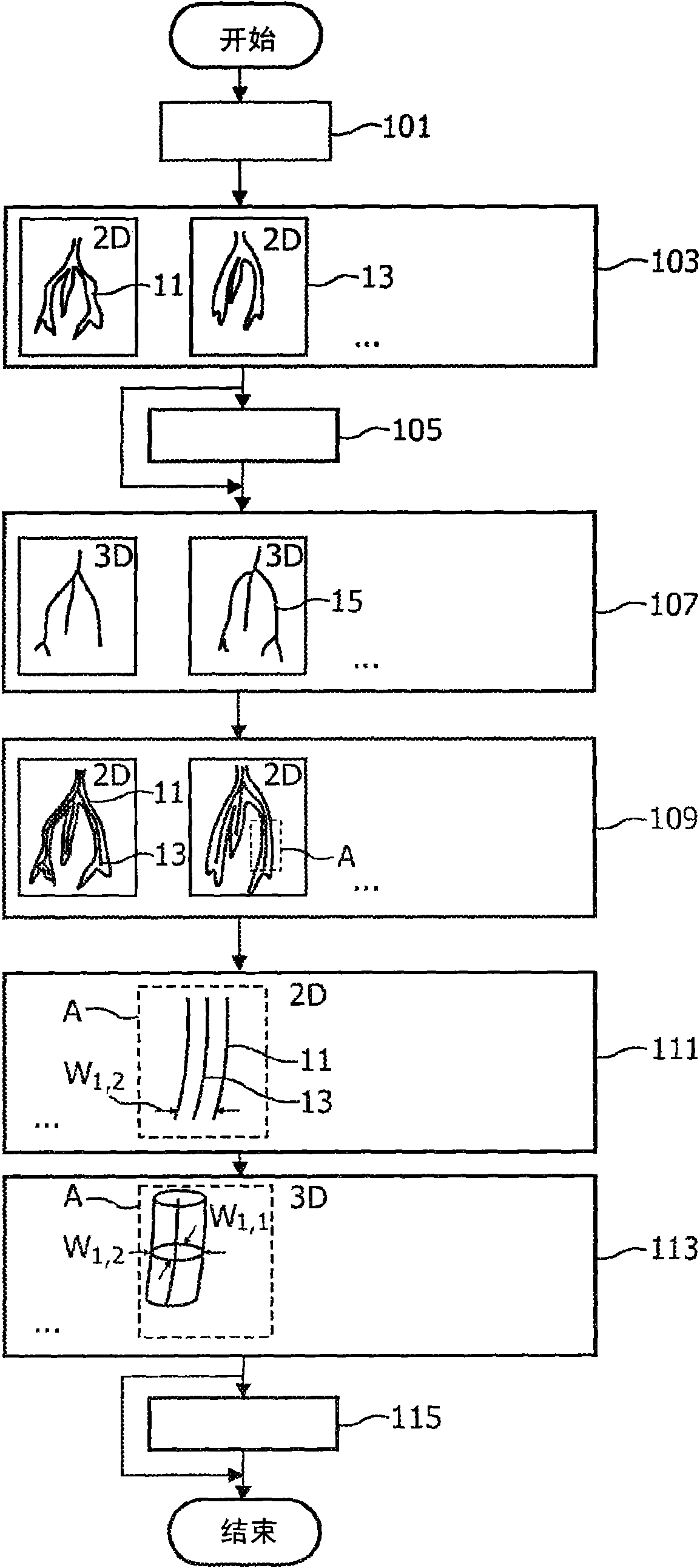
[0035] can use figure 1 Basic steps of a method for acquiring a 3-dimensional image of a coronary vein according to an embodiment of the present invention are explained.

[0036] After positioning the patient in a suitable device, such as a C-arm X-ray device, a catheter is used to inject contrast medium into the coronary veins to be imaged (step 101 ).

[0037] Then, while rotating the C-arm around the patient's torso, multiple 2D X-ray images of the viewing area including the vein 11 are acquired at different projection angles (step 103) (only two images 13 are shown exemplary ).

[0038] Optionally, the acquired 2D image may be down-sampled and / or filtered (step 105) using a high-pass filter and / or vessel enhancement filter, thereby improving image quality for the vein to be imaged.

[0039] A 3D midline model 15 of the venous system is derived (step 107 ) from a certain number of 2D images acquired during the same motion phase (eg end-diastole where cardiac motion is min...


PUM


Abstract

A method and an apparatus for acquiring 3-dimensional images of coronary vessels (11), particularly of coronary veins, is proposed. 2-dimensional X-ray images (13) are acquired within a same phase ofa cardiac motion. Then, a 3- dimensional centerline model (15) is generated based on these 2-dimensional images. From 2-dimensional projections of the centerline model into respective projection planes, the local diameters (w) of the vessels in the projection plane can be derived. Having the diameters, a 3-dimensional hull model of the vessel system can be generated and, optionally, 4-dimensionalinformation about the vessel movement can be derived.

Description

technical field [0001] The invention relates to a method for acquiring a 3-dimensional image of a coronary vessel, especially a 3-dimensional image of a moving coronary vein in a periodic motion. Furthermore, the invention relates to a device adapted to perform such a method, a computer program adapted to perform such a method when run on a computer, and a computer-readable medium comprising such a program. Background technique [0002] For medical purposes, it may be important to know precisely the position, size, shape and / or motion of coronary vessels. For example, for a surgical treatment such as implanting a stent into a coronary vessel, the surgeon must know the geometry of the vasculature to be treated, the location where the stent is to be placed, and preferably also the motion of the vasculature during the surgical procedure. Therefore, it may be advantageous to provide a 3-dimensional image of the vasculature to be treated so that the surgeon can analyze the surgi...

Claims


Application Information

Patent Timeline
Patent Type & Authority Applications(China)
IPC IPC(8): A61B6/00G06T11/00
CPCA61B6/463G06T2207/10116A61B6/481G06T17/00G06T7/0012G06T2200/08G06T11/006A61B6/504G06T2207/10112A61B6/541G06T2207/20044G06T7/60A61B6/4441G06T2211/404G06T7/0067G06T7/0077G06T2207/30101G06T2211/412G06T7/564G06T7/596
Inventor U·扬特D·舍费尔M·格拉斯
Owner KONINK PHILIPS ELECTRONICS NV
Why Eureka
Try Eureka