Articular cartilage restoration and regeneration stent and preparation method thereof

A technology for repairing and regenerating articular cartilage, applied in the field of medical materials, it can solve the problems of poor biocompatibility of artificial scaffold materials, affecting cell growth, poor mechanical mechanics, etc., achieving sufficient and safe donor sources, satisfying clinical application, and easy clinical practice. popular effect

Active Publication Date: 2013-10-09
BEIJING WANJIE MEDICAL DEVICE CO LTD
View PDF7 Cites 1 Cited by
  • Summary
  • Abstract
  • Description
  • Claims
  • Application Information

AI Technical Summary

Problems solved by technology

[0004] In the process of realizing the present invention, the inventors found that the prior art has at least the following problems: the material properties of natural scaffold materials cannot be changed, and the mechanical strength and degradation rate are difficult to control, etc.
Commonly used natural scaffold materials include collagen, hydroxyapatite, etc. The biomechanical properties of natural scaffolds are difficult to adjust, and it is difficult to find biomechanical properties similar to those of cartilage.
Some natural biomaterial scaffolds have poor mechanical mechanics and cannot resist the mechanical stress generated during joint movement, which may easily cause the repair site to collapse and also affect the growth of cells on the material. For example, the elasticity and creep of hydroxyapatite scaffolds are higher than those of cartilage. Poor, too high hardness can easily cause wear of the corresponding cartilage articular surface
The biocompatibility of artificial scaffold materials is relatively poor, and there are different degrees of inflammatory reactions, as well as immunogenicity problems, and even carcinogenicity problems

Method used


Image

  • Articular cartilage restoration and regeneration stent and preparation method thereof
  • Articular cartilage restoration and regeneration stent and preparation method thereof
  • Articular cartilage restoration and regeneration stent and preparation method thereof

Examples


Embodiment 1

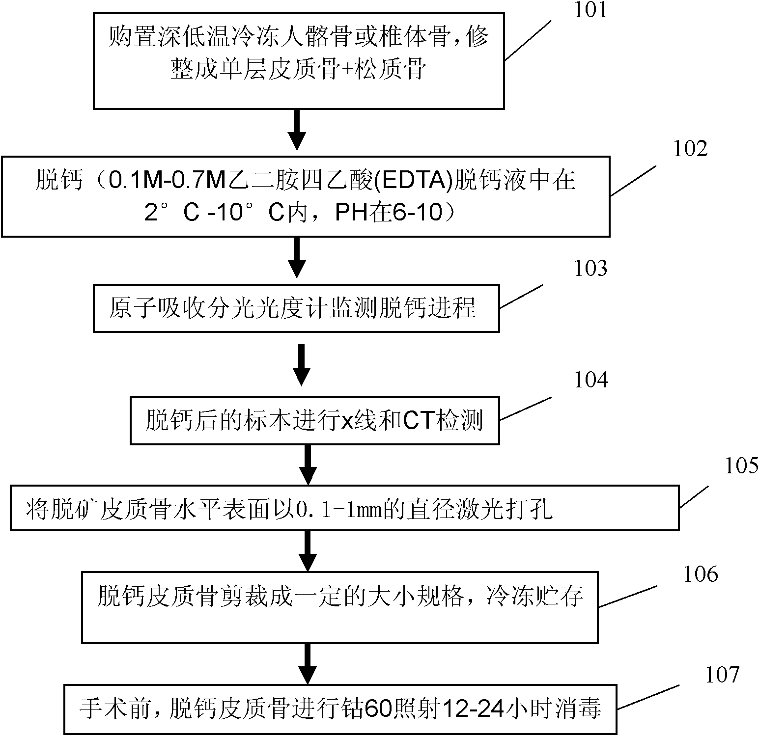
[0043] see image 3 , a method for preparing a scaffold for articular cartilage repair and regeneration, comprising the following steps:

[0044]Step 101: Select the bone with cancellous bone under the cortical bone, trim the thickness of the cortical bone to 0.5-3mm, decalcify the solution as 0.1M-0.7M EDTA, pH 6-10, temperature: 2-10 ℃;

[0045] Step 102: Soak the bones in a decalcification solution for decalcification;

[0046] Step 103: monitoring the decalcification process with an atomic absorption spectrophotometer;

[0047] Step 104: Carry out x-ray and CT detection on the decalcified specimen to prove that the bone is completely decalcified to obtain decalcified cancellous-cortical bone;

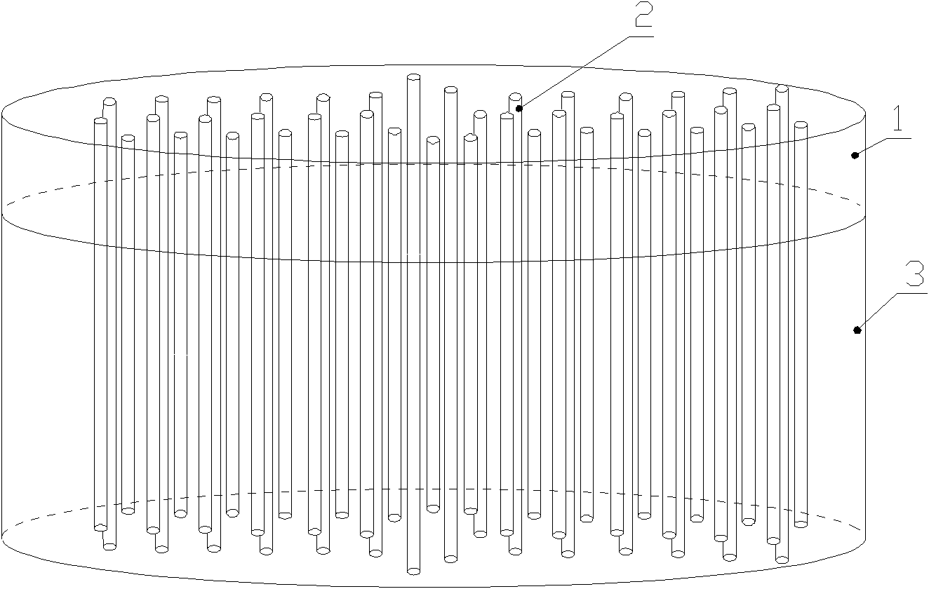
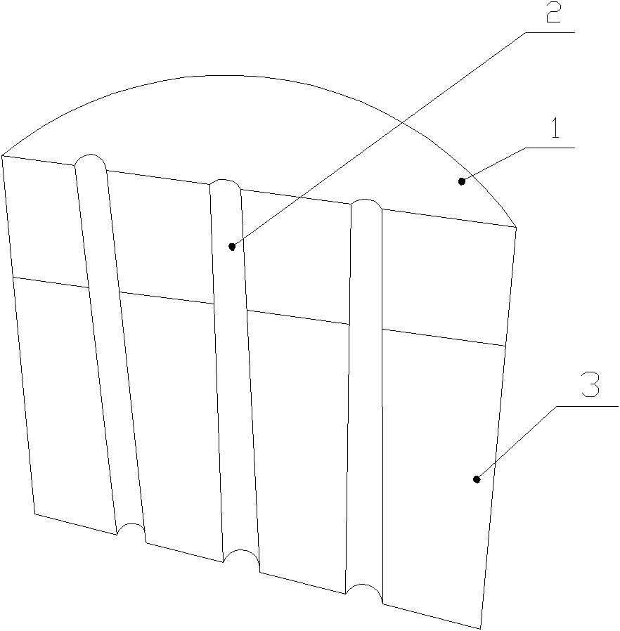
[0048] Step 105: Laser drilling holes with a diameter of 0.1-1 mm on the horizontal surface of the decalcified cortical bone;

[0049] Step 106: Detect the nano-elastic modulus of each part of the decalcified cancellous-cortical bone scaffold with a nano-indentation biomechanica...

Embodiment 2

[0055] Preparation method of scaffold for articular cartilage repair and regeneration:

[0056] The preparation flow chart is as image 3 As shown, purchase cryopreserved human ilium or vertebral bone, soak in 0.1M-1 MEDTA decalcification solution, the temperature of the decalcification solution is within 6°C, and the pH is 8, and the cortical bone is removed by chelation. Calcium, decalcification solution was changed daily. Prepare EDTA decalcification solution with deionized water and store it in a plastic container. Use an atomic absorption spectrophotometer to detect the calcium ion concentration in the decalcification solution that is changed every day to monitor the decalcification process. When the Ca chelate concentration in the decalcification solution is figure 2 As shown, the horizontal surface of the decalcified cortical bone is drilled vertically with a diameter of 0.5mm, and the drilling is done by laser drilling. Therefore, the hole diameter and hole distance...


PUM

PropertyMeasurementUnit
diameteraaaaaaaaaa
distanceaaaaaaaaaa
porosityaaaaaaaaaa

Abstract

The invention discloses an articular cartilage restoration and regeneration stent and a preparation method thereof, belonging to the field of medical materials. The stent is made of decalcified spongiosa-cortical bone, and the decalcified spongiosa-cortical bone is provided with more than two holes penetrating through the decalcified spongiosa bone and the cortical bone. The holes are vertically arranged on the horizontal surface of the decalcified cortical bone and have diameter of 0.1-1mm. The width between hole centers is 0.5-5mm. The preparation method provided by the invention comprises the steps of: soaking the bone in a decalcified solution for decalcification; monitoring the decalcification process by using an atomic absorption spectrophotometer; carrying out X-ray and CT detection on a decalcified sample to prove that the bone is completely decalcified to obtain the decalcified spongiosa-cortical bone; drilling on the horizontal surface of the decalcified cortical bone by using laser in a diameter of 0.1-1mm; and cutting the decalcified cortical bone into a certain size, and freezing and storing. The articular cartilage restoration and regeneration stent is made of a natural collagen stent material without immunological rejection reaction, is easy to adsorb by cells, and has good biocompatibility and better mechanical strength.

Description

technical field [0001] The invention relates to the field of medical materials, in particular to a scaffold for articular cartilage repair and regeneration and a preparation method thereof. Background technique [0002] Articular cartilage injury is an intractable injury in the field of sports medicine. The incidence of articular cartilage injury caused by exercise is very high. According to the survey results of the Institute of Sports Medicine of Peking University, cartilage injury accounts for more than 40.5% of knee joint sports injuries. Cartilage damage often causes symptoms such as joint swelling and pain. Due to the special structure of cartilage tissue, there are no nerves, blood vessels and lymphatic vessels in it, and the nutrient supply of cartilage is through the pressure changes generated by joint activities, so that the synovial fluid communicates between the joint cavity and the cartilage matrix, so the damaged articular cartilage cannot Repair and regenera...

Claims


Application Information

Patent Timeline
Patent Type & Authority Patents(China)
IPC IPC(8): A61L27/36A61L27/56
Inventor 敖英芳张辛何震明马勇周春燕
Owner BEIJING WANJIE MEDICAL DEVICE CO LTD
Why Eureka
Try Eureka