Bracket for regenerating decalcification cortical-bone articular cartilage with vertical drill and preparation method thereof

A technology of articular cartilage and cortical bone, used in joint implants, joint implants, bone implants, etc., can solve the problems of biocompatibility and mechanical strength being difficult to take into account at the same time, complicated operation, etc., and achieve easy clinical Effects of application, enhanced repair ability, increased mechanical strength

Active Publication Date: 2011-07-20
PEKING UNIV THIRD HOSPITAL
View PDF0 Cites 0 Cited by
  • Summary
  • Abstract
  • Description
  • Claims
  • Application Information

AI Technical Summary

Problems solved by technology

[0004] To sum up, the above-mentioned methods have more or less shortcomings in that it is difficult to balance biocompatibility and mechanical strength at the same time, and most of them need to be cultured in vitro, which makes the operation complicated.

Method used


Image

  • Bracket for regenerating decalcification cortical-bone articular cartilage with vertical drill and preparation method thereof
  • Bracket for regenerating decalcification cortical-bone articular cartilage with vertical drill and preparation method thereof
  • Bracket for regenerating decalcification cortical-bone articular cartilage with vertical drill and preparation method thereof

Examples


Embodiment 1

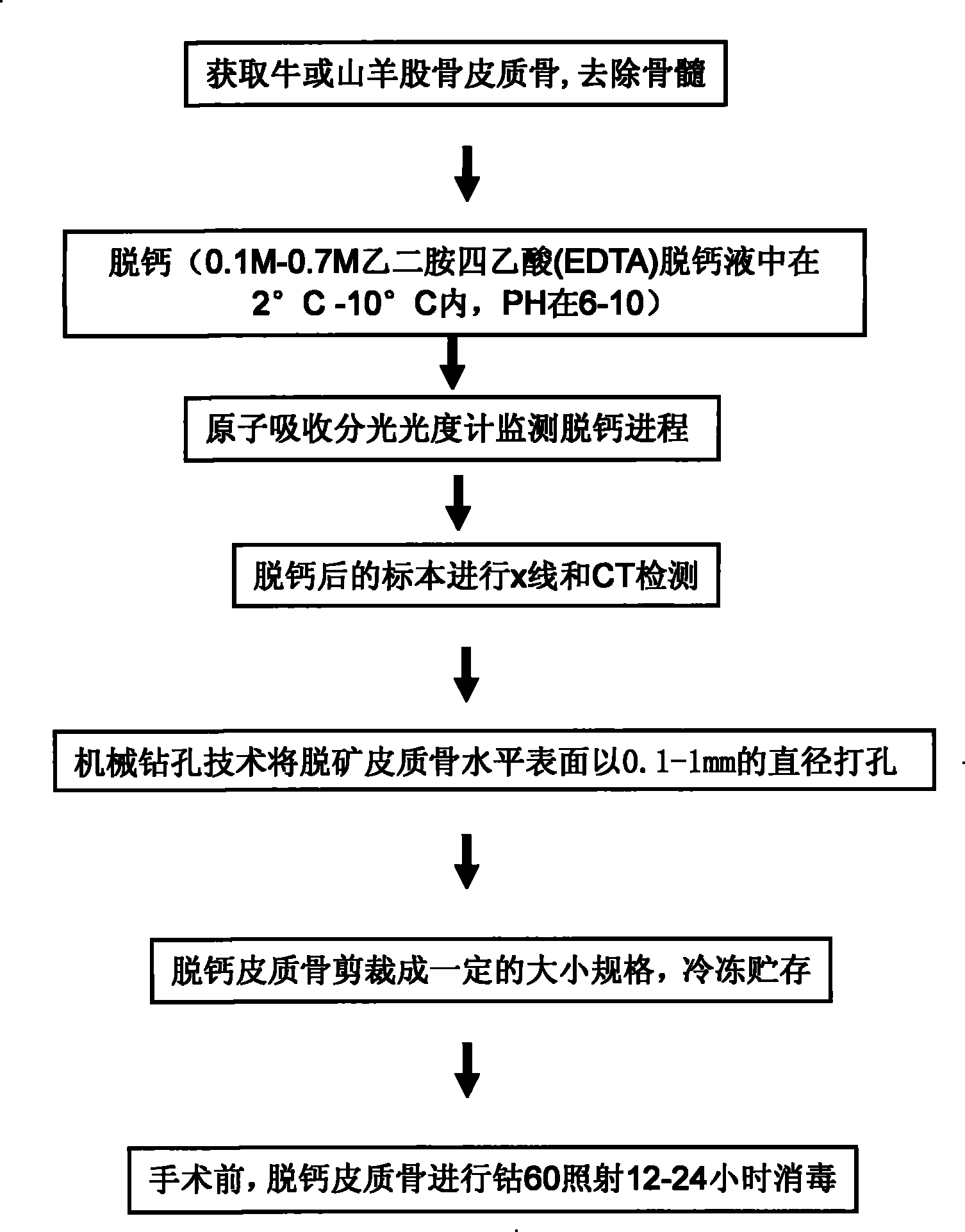
[0036] Preparation method of longitudinally drilled decalcified cortical bone scaffold for articular cartilage regeneration

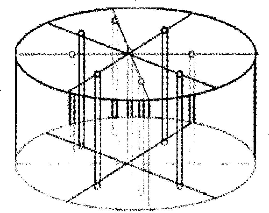
[0037] For the preparation flow chart, see figure 1 , Take the femoral backbone of cattle or goats, remove the bone marrow, soak in 0.1M-0.7M ethylenediaminetetraacetic acid (EDTA) decalcification solution at 2°C-10°C, pH between 6-10, use chelation The calcium in the cortical bone is removed, and the decalcification solution is replaced daily. Prepare EDTA decalcification solution with deionized water and store it in plastic containers (avoid glass containers). Use an atomic absorption spectrophotometer to detect the calcium ion concentration in the decalcification solution that is changed every day to monitor the decalcification process. When the Ca chelate concentration in the decalcification solution is image 3 As shown, the horizontal surface of the decalcified cortical bone is vertically drilled with a diameter of 0.1-1mm (the aperture in the ex...

Embodiment 2

[0041] Experimental procedure of repairing cartilage defect in rabbit animal model

[0042] In order to verify the effect of the longitudinally drilled decalcified cortical bone articular cartilage regeneration scaffold in the repair method, we conducted a simulated operation experiment on a rabbit animal model. Select 2.5-3.0kg New Zealand big-eared white rabbits, anesthetize, prepare skin, and disinfect, such as Figure 6 As shown, the incision of the knee joint exposes the femoral trochlear, and a 4mm diameter and 2mm deep defect is made on the femoral trochlea. After microfracture treatment in the subchondral bone drilling, the 4mm diameter of the decalcified cortical bone scaffold is removed in the sterilized drill hole. 2 mm high cylinder, filled into the defect. The wound was sutured in layers, and samples were taken 12 weeks after the operation for nuclear magnetic resonance, electron microscopy, and histological observations. The results are shown in Figure 7-9 . ...


PUM

PropertyMeasurementUnit
thicknessaaaaaaaaaa
porosityaaaaaaaaaa
porosityaaaaaaaaaa

Abstract

The invention discloses a stent used for the joint cartilage regeneration of the decalcification cortex bone of longitudinal drilling and a preparation method thereof, and the stent belongs to an animal decalcification cortex bone. Holes with the diameter of 0.1 to 1 mm are drilled perpendicularly on the horizontal surface of the decalcification cortex bone; the interval among the holes is 0.5 to5 mm, thereby forming three-dimensional porous stereo network structure; the porosity reaches 5 percent to 20 percent. The preparation method is as follows: a first step, the decalcification of the cortex bone: a cortex bone is taken out from the cattle or goat to remove the bone marrow and then soaked in decalcifying solution which is replaced everyday and the cortex bone is completely decalcified to enter into the next step; a second step, drilling: holes with the diameter of 0.1 to 1 mm are drilled on the horizontal surface of the decalcification cortex bone; the interval among the holes is supposed to be 0.5 to 5 mm, after the cortex bone is tailored into a designed dimension, then a stent used for the joint cartilage regeneration of the decalcification cortex bone of longitudinal drilling is gotten. The stent used for the joint cartilage regeneration of the decalcification cortex bone of longitudinal drilling has extensive material selection and quite good biocompatibility.

Description

technical field [0001] The invention belongs to the field of preparation of artificial articular cartilage, and in particular relates to utilizing existing biological materials to prepare artificial longitudinally drilled decalcified cortical bone articular cartilage regeneration scaffold and a preparation method thereof. Background technique [0002] The biological research of articular cartilage has confirmed that due to the special structure of cartilage tissue, there are no nerves, blood vessels, and lymphatic vessels in it. Although juvenile articular cartilage can obtain part of the nutrition through the subchondral pathway, adult articular cartilage has no subchondral nutrition pathway. The nutrition of cartilage The supply is through the pressure changes generated by joint activities, so that the synovial fluid communicates between the joint cavity and the cartilage matrix, so the cartilage itself lacks effective repair ability, and the treatment of injuries is someti...

Claims


Application Information

Patent Timeline
Patent Type & Authority Patents(China)
IPC IPC(8): A61L27/36A61F2/30A61F2/28A61B17/56
Inventor 敖英芳张辛周春燕刘平马勇
Owner PEKING UNIV THIRD HOSPITAL
Why Eureka
Try Eureka