Method for preparing amniotic compound corneal limbus stem cell membrane

A technology of corneal limbal stem cells and cell membranes, which is applied in the field of preparation of amniotic membrane composite corneal limbal stem cell membranes, can solve the problems of uneven layering of cultured epithelium, loss of stem cell numbers, and effect effects, and achieve uniform layering of limbal stem cells Effect

Active Publication Date: 2011-08-31
SHANDONG EYE INST
View PDF5 Cites 8 Cited by
  • Summary
  • Abstract
  • Description
  • Claims
  • Application Information

AI Technical Summary

Problems solved by technology

At present, there are mainly two methods of culturing limbal stem cells commonly used for transplantation. One is to flatten the amniotic membrane on the surface of the cell culture vessel or glass slide, and the obtained cell membrane needs to be removed from the culture vessel before transplantation. It is very easy to expose and affect clinical use. At the same time, this method cannot be co-cultured with feeder cells, which wil...

Method used


Image

  • Method for preparing amniotic compound corneal limbus stem cell membrane
  • Method for preparing amniotic compound corneal limbus stem cell membrane

Examples


preparation example Construction

[0012] Preparation method of the present invention:

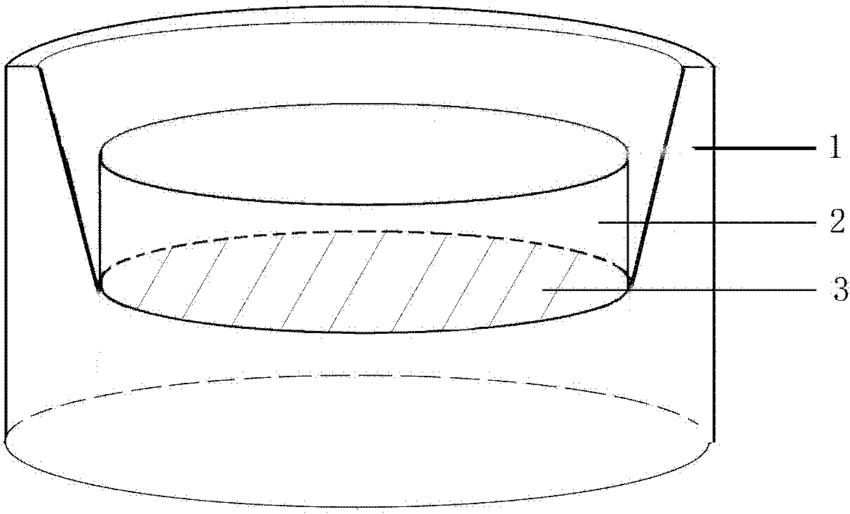
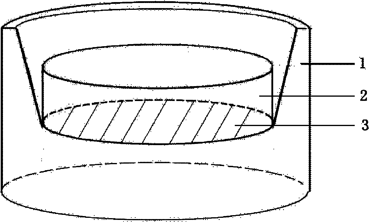
[0013] 1. Preparation of amniotic membrane nested culture mold:

[0014] Such as figure 1 , according to the conventional method to prepare 5.0cm × 5.0cm size of sterile amniotic membrane 3 amniotic membrane epithelium, the amniotic membrane 3 epithelial side up in a 6-well culture plate; On the end face of the sleeve 2 made of polypropylene material, the embedded culture chamber 1 is buckled on the amniotic membrane sheet 3 of the sleeve 2 to form an amniotic membrane nested culture mold. Wherein used sleeve 2 external diameter 24mm, high 16mm.

[0015] Two: Preparation of mouse embryonic fibroblast feeder layer:

[0016] Treat adherent mouse embryonic fibroblasts with 0.01mg / ml mitomycin C solution and incubate at 37°C for 2 hours; wash the cells carefully with 10ml PBS phosphate buffer; add 1ml 0.25% trypsin / 0.02% EDTA , incubate at 37°C for 3-5 minutes; add 9ml of serum-containing DMEM medium to stop digestion, pipe...

Embodiment 1

[0021] Prepare the epithelium-free sterile amniotic membrane sheet 3 with a size of 5.0cm×5.0cm according to the conventional method, spread the epithelial surface of the amniotic membrane sheet 3 on a 6-well culture plate; The end face of the sleeve 2 with an outer diameter of 24 mm and a height of 16 mm made of material is buckled on the amnion sheet 3 of the sleeve 2 with an embedded culture chamber 1 to form an amnion nested culture mold. A feeder layer of mouse embryonic fibroblasts was prepared in the wells of a 6-well plate according to a conventional method, and an amniotic membrane nested culture mold was placed on the feeder layer. Take two remaining fresh corneal rings from the eye bank, soak them in DMEM culture solution containing 100 units / ml penicillin and streptomycin antibiotics, rinse them, and then put them into DMEM culture solution containing 2.4 units / ml neutral protease Digest in a petri dish at 37°C for 1 hour; remove the tissue pieces from neutral prot...


PUM


Abstract

The invention relates to a method for preparing an amniotic compound corneal limbus stem cell membrane, which comprises the following steps of: firstly, preparing a sterile amniotic membrane with epithelium removed, placing the epithelial surface of the amniotic membrane downwards on an end face of a sleeve made of a poly propylene material, and covering an embedded culture transwell on the amniotic membrane of the sleeve to obtain an amniotic-membrane-embedded culture mold; placing the culture mold in holes in a 6-hole plate on which a mouse embryonic fibroblast cell feeder layer is spread; and finally, preparing a corneal limbus stem cell suspension by using a digestion method, inoculating the suspension on the epithelial surface of the amniotic membrane, inoculating 2.0-3.0*10<5> corneal limbus stem cells for each culture mold, and culturing the corneal limbus stem cells for 10 days to obtain the amniotic compound corneal limbus stem cell membrane. Compared with the traditional amniotic membrane spreading method and the latex ring amniotic membrane fixing method, the amniotic compound corneal limbus stem cell membrane prepared with the method has flat amniotic membrane culturing surface and uniform corneal limbus stem cell stratified layer and especially has the advantage of convenience for clinic application and difficulty in rapture of the amniotic membrane.

Description

technical field [0001] The invention belongs to the field of corneal limbal stem cell culture, and in particular relates to a preparation method of amniotic membrane composite corneal limbal stem cell membrane. Background technique [0002] Corneal epithelial cells come from stem cells in the limbus, and the normal limbus is the key to maintaining normal ocular surface function. After the corneal limbus is damaged, the function defect or insufficient number of stem cells in the damaged area will cause the conjunctival epithelium to grow into the corneal surface, and corneal neovascularization will further cause severe symptoms such as corneal scarring, resulting in a significant decrease in vision or even loss of vision. Supplementing a certain amount of stem cells through transplantation is currently one of the most effective treatments. [0003] Limbal stem cell transplantation has become a research hotspot of many scholars due to limitations of material sources and posto...

Claims


Application Information

Patent Timeline
IPC IPC(8): A61L27/38C12N5/074
Inventor 周庆军史伟云谢立信王瑶杨玲玲
Owner SHANDONG EYE INST
Why Eureka
Try Eureka