
Schistosoma japonicum 23kDa membrane protein big hydrophilic peptide segment fusion protein and application thereof in schistosome infection immune diagnosis

A fusion protein, schistosomiasis technology, applied in the direction of DNA/RNA fragments, peptides, hybrid peptides, etc., can solve the problems of insufficient specificity and early diagnostic value, achieve good diagnostic value of schistosomiasis, good application prospects, and improve sensitivity with specific effects

Inactive Publication Date: 2011-08-31
JIANGSU INST OF PARASITIC DISEASES +1
View PDF1 Cites 0 Cited by
  • Summary
  • Abstract
  • Description
  • Claims
  • Application Information

AI Technical Summary

Problems solved by technology

[0013] The purpose of the present invention is to provide a recombinant fusion protein as a detection antigen to detect anti-Schistosoma japonicum 23kDa membrane protein molecular specific antibody IgG or IgM immunoblotting method in the host serum, which can solve the lack of specificity and early diagnosis value of the current schistosomiasis antibody detection method The problem

Method used


Image

  • Schistosoma japonicum 23kDa membrane protein big hydrophilic peptide segment fusion protein and application thereof in schistosome infection immune diagnosis
  • Schistosoma japonicum 23kDa membrane protein big hydrophilic peptide segment fusion protein and application thereof in schistosome infection immune diagnosis
  • Schistosoma japonicum 23kDa membrane protein big hydrophilic peptide segment fusion protein and application thereof in schistosome infection immune diagnosis

Examples


Embodiment 1

[0067] Embodiment 1: human serum albumin gene cloning

[0068] The human serum albumin gene is reverse-transcribed and synthesized from the mRNA of human fetal liver tissue by RT-PCR technology. The specific preparation method is as follows:

[0069] 1. Preparation of human fetal liver mRNA

[0070] Take 0.5 g of freshly isolated human fetal liver tissue, freeze it in liquid nitrogen, pulverize it in a ceramic mortar, and use the mRNA purification kit (Illustra QuickPrep TM mRNA purification kit) was used to prepare and purify mRNA, and the operation method was strictly in accordance with the operation instructions of the kit. The purity and content of mRNA were measured by UV spectrophotometer.

[0071] 2. HSA gene amplification

[0072] 2.1 Primer design:

[0073] HSA1: 5′-GATGCACACAAGAGTGAGGT-3′

[0074] HSA2: 5'-AACTCGAGTTATAAGCCTAAGGCAGCTTGACTTGC-3'.

[0075] 2.2 First strand cDNA synthesis

[0076] Using Phusion TM RT-PCR Kit (purchased from NEB Beijing Branch)...

Embodiment 2

[0079] Example 2: Sj23HD gene synthesis

[0080] The Sj23HD gene is prepared by artificial synthesis. In order to facilitate the fusion of the Sj23HD gene and the HSA gene, and to facilitate gene cloning, a Nco 1, the 5' end DNA sequence of part of the HSA gene is carried at its 3' end. The synthesized Sj23HD gene is shown as SEQ ID NO:5. The synthesis of the gene sequence was completed by Shanghai Handsome Biotechnology Co., Ltd.

Embodiment 3

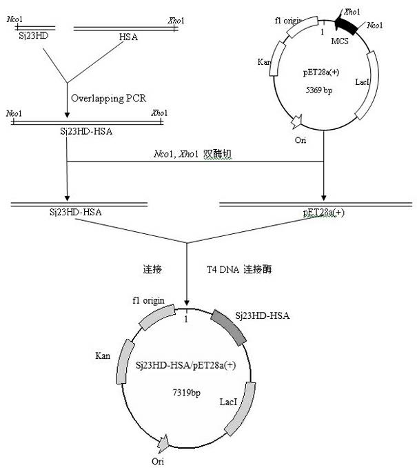
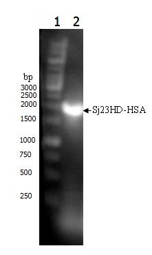
[0081] Example 3: Construction and sequence analysis of Sj23HD-HSA fusion protein gene

[0082] The preparation of the Sj23HD-HSA fusion protein gene is carried out according to the principle of Overlapping PCR.

[0083] Primer design:

[0084] Sj23HD1: 5′-CATGGATGACTGGTGCTCT-3′,

[0085] HSA2: 5'-AACTCGAGTTATAAGCCTAAGGCAGCTTGACTTGC-3'.

[0086] Gene Amplification:

[0087] In a 0.2mL PCR tube, add 2×Phusion Master Mix 25μL, synthetic Sj23HD gene fragment 2μL, purified HSA gene fragment 2μL (the molecular ratio of these two gene fragments should be adjusted to 1:1 as much as possible), add no Ionized water to a final volume of 48 μL, 98°C for 30 Sec; anneal at 65°C for 30 Sec, extend at 72°C for 2 min, and cool the reaction tube on ice. Add 1 μL each of primers Sj23HD1 and HSA2, mix well, and centrifuge to collect the reaction product at the bottom of the tube. Then carry out gene amplification according to the following conditions: 98°C, 10 Sec; 65°C, 20 Sec, 72°C, 50 Sec,...


PUM


Abstract

The invention relates to schistosoma japonicum 23kDa membrane protein big hydrophilic peptide segment fusion protein and application thereof in schistosome infection immune diagnosis and belongs to the technical field of immunological diagnosis of parasitic diseases. The invention relates to fusion protein of schistosoma japonicum 23kDa membrane protein big hydrophilic peptide segment (Sj23HD) and human serum albumin (HAS). The fusion protein comprises two polypeptide regions, wherein the first region is the Sj23HD; the second region is the HAS; the C tail end of the first region is directly connected with the N tail end of the second region without any connecting peptide; and the structural formula is Sj23HD-HAS. The Sj23HD-HAS fusion protein remains the reactogenicity of the Sj23HD and has higher stability. The Sj23HD-HAS fusion protein, serving as a detecting antigen to diagnose the schistosome infection by the Western blot, has higher sensitivity and specificity.

Description

technical field [0001] The invention relates to preparation of 23kDa membrane protein large hydrophilic peptide fusion protein antigen of Schistosoma japonicum and a kit and detection method for early diagnosis of Schistosoma infection based on immunoblotting method, belonging to the technical field of parasitic disease immunodiagnosis. Background technique [0002] Schistosomiasis is a serious public health problem that seriously endangers the health of people in tropical and subtropical regions of the world. There are still 449 counties in China where schistosomiasis is prevalent, nearly 440,000 schistosomiasis patients, and about 230 million people are threatened by schistosomiasis infection. The key measures for the prevention and treatment of schistosomiasis are to diagnose the source of infection such as patients, domestic animals and wild animals infected by schistosomiasis, give timely treatment, eliminate the source of infection, and block the spread of schistosomia...

Claims


Application Information

Patent Timeline
Patent Type & Authority Applications(China)
IPC IPC(8): C07K19/00C12N15/62C12N15/70G01N33/53
Inventor 余传信王玠张伟钱春艳宋丽君殷旭仁梁幼生高琪
Owner JIANGSU INST OF PARASITIC DISEASES
Learn More