Zoom multi-channel microscopic imaging system of eye retina

A microscopic imaging, multi-channel technology, applied in the field of human imaging, can solve the problems of fast switching of light source parameters, limited exposure time and imaging frame rate of serial imaging, neglect of spectral information and polarization information of retinal tissue, etc.

Active Publication Date: 2011-09-21
INST OF OPTICS & ELECTRONICS - CHINESE ACAD OF SCI
View PDF8 Cites 10 Cited by
  • Summary
  • Abstract
  • Description
  • Claims
  • Application Information

AI Technical Summary

Problems solved by technology

[0005] (1) Ignoring the rich spectral information and polarization information of retinal tissue, which is of great value for characterizing tissue physiological parameters;
[0006] (2) When it is necessary to observe a variety of microstructures (such as three-color cone cells, capillaries, etc.), since the targets have different reflectivity and absorption characteristics, it is usually unpredictable, so a single illumination imaging wavelength cannot Optimal image contrast;
[0007] (3) Although some people use the method of time-sharing lighting and post-synthesis as an improved solution, it is relatively troublesome to quickly switch the light source parameters, and it is difficult to control the volume and complexity of the system; in addition, more importantly, under various lighting conditions Serial imaging is limited by the exposure time and imaging frame rate (at present, it can only reach tens of Hz at most), so it is difficult to avoid the difficulty of image registration caused by the inherent physiological vibration of the retina. Correlation, therefore, this scheme is not satisfactory in terms of quality or efficiency in actual use

Method used


Image

  • Zoom multi-channel microscopic imaging system of eye retina
  • Zoom multi-channel microscopic imaging system of eye retina
  • Zoom multi-channel microscopic imaging system of eye retina

Examples


Embodiment Construction

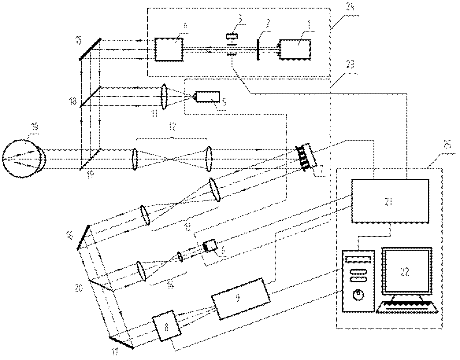
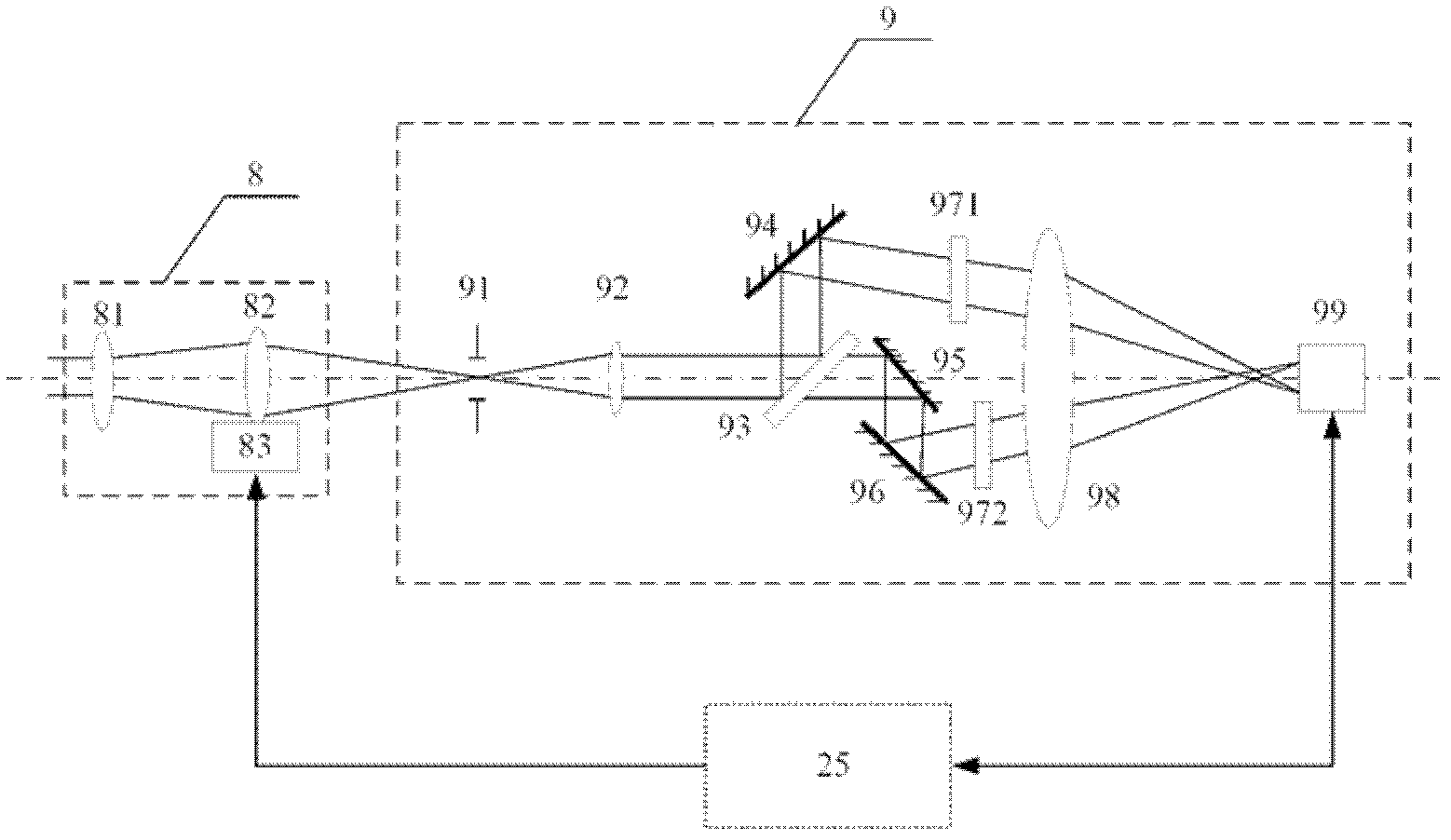
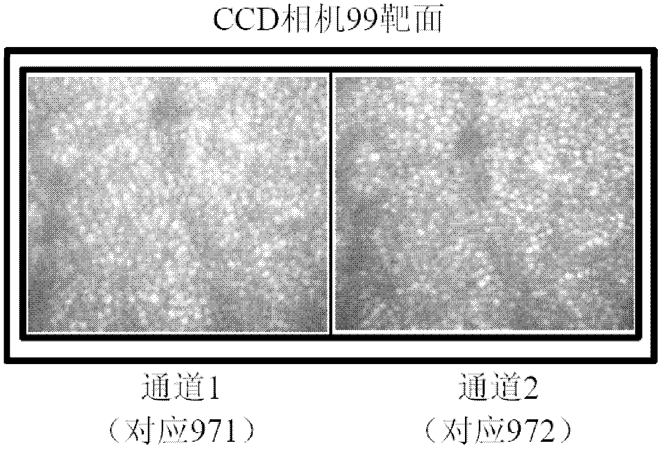
[0028] Such as figure 1 As shown, a zoom multi-channel living human retina microscopic imaging system according to an embodiment of the present invention includes an illumination subsystem 24, a zoom module 8, a multi-channel imaging module 9 and a control device composed of a system circuit 21 and a PC 22 25, shown by the dashed box in the figure.

[0029] In the present invention, the lighting subsystem 24 emits pulsed lighting light under the control of the control device 25 . To this end, the illumination subsystem 24 may include an illumination source 1 that emits continuous light, and an optical switch 3 that modulates the light emitted by the illumination source 1 to form pulsed light. If the illumination light source 1 itself is a pulsed light source, the optical switch 3 is not necessary.

[0030] Optionally, the lighting subsystem 24 may further include a filter 2 for performing spectral selective filtering on the light emitted by the lighting source 1 . If the il...


PUM


Abstract

The invention discloses a zoom multi-channel microscopic imaging system of eye retina, which comprises a lighting subsystem, a zooming module, a multi-channel imaging module and a controller, wherein the lighting subsystem is used for emitting light to light eye ground, and the light is reflected by retina tissues and then forms imaging light beams; the zooming module is suitable for optical zooming so as to focus-image all the depth zones of the retina and lead to different magnifying powers and fields of view; the multi-channel imaging module is used for filtering and arranging the imaging beams spatially after the imaging beams are split and finally focusing the light on the target surface of a photoelectric detector to form a plurality of twin images; and the controller is used for connecting and controlling all the other parts, taking pictures of the eye ground of the tested person continuously, and collecting and obtaining a video or an image sequence directly. The zoom multi-channel microscopic imaging system also comprises a self-adaptive optical subsystem which is used for correcting higher order aberration of human eyes in real time so as to reach an optical resolution ratio approaching the diffraction limit and realize microscopic observation of the living eye retina.

Description

technical field [0001] The invention belongs to the technical field of human body imaging, and in particular relates to a high-resolution microscopic imaging device for living human eye retina, especially a high-resolution living human eye retina high-resolution microscopic imaging device characterized by multi-channel continuous dynamic imaging and supplemented with optical zoom function. rate microscopy imaging system. Background technique [0002] Retinal imaging is of great value for medical research and clinical diagnosis. Previously, digital fundus cameras, confocal scanning ophthalmoscopes and other hospital ophthalmology equipment can only acquire retinal images with a large field of view, while the resolution of in vivo retinal microscopic imaging is limited by the aberrations of the human eye's refractive system, especially high-order images. However, no substantial progress has been made. In 1997, Liang, David Williams and others corrected the wavefront aberrati...

Claims


Application Information

Patent Timeline
Patent Type & Authority Applications(China)
IPC IPC(8): A61B3/12A61B3/15
Inventor 杨汉生张雨东饶学军戴云
Owner INST OF OPTICS & ELECTRONICS - CHINESE ACAD OF SCI
Why Eureka
Try Eureka