Time-resolved immunoflourometric chromatographic test strip for quantitative detection as well as preparation method and application thereof

A technology for time-resolved fluorescence and detection of test paper, which is applied in the direction of analytical materials, measuring devices, instruments, etc., can solve the problems of complex production process, high requirements for instruments and equipment, and difficulty in fixing antibodies, and achieve the effect of high sensitivity

Active Publication Date: 2011-09-21
BEYOND DIAGNOSTICS (SHANGHAI) CO LTD
View PDF4 Cites 61 Cited by
  • Summary
  • Abstract
  • Description
  • Claims
  • Application Information

AI Technical Summary

Problems solved by technology

Whatman, a subsidiary of GE, has developed Fusion5, a membrane with five-in-one functions, which combines the functions of the above five membranes. However, due to the large pore size of Fusion5, it is difficult to immobilize antibodies. It needs to prepare special Stone for immobilizing antibodies, and the production process is relatively complicated. The requirements for instruments and equipment are relatively high, and the developed immunochromatographic reagents can only be used for qualitative detection

Method used


Image

  • Time-resolved immunoflourometric chromatographic test strip for quantitative detection as well as preparation method and application thereof
  • Time-resolved immunoflourometric chromatographic test strip for quantitative detection as well as preparation method and application thereof
  • Time-resolved immunoflourometric chromatographic test strip for quantitative detection as well as preparation method and application thereof

Examples


Embodiment 1

[0041] Example 1 Preparation of quality control microspheres coated with biotin-labeled γ-globulin (BGG)

[0042] (1) Preparation of biotinylated BGG

[0043] with 0.1M NaCNBH 3BGG (purchased from Pel-Freez Biological) was formulated into a 10mg / ml solution, and Biotin-X-X-NHS (N-hydroxysuccinimide modified biotin) was prepared using DMSO (dimethylformamide), manufacturer: SIGMA, product No.: B3295) solution to 16.172mg / ml, add the Biotin-X-X-NHS solution to the BGG solution according to the amount of 1mg antibody added to 5.4ul Biotin-X-X-NHS, mix well and place overnight at 4°C. Dialysis was used to remove free unreacted biotin, and the dialysate was biotin-labeled antibody dialysate buffer (0.1M Tris, 0.3MNaCl, 0.005MEDTA-Na-2H 2 O, pH8.0). After dialysis, the protein concentration was determined by BCA method.

[0044] (2) The luminescent microspheres coated with aldehyde group modified by the above-mentioned labeled protein

[0045] Add 6.4 μl of 20% Tween-20 solutio...

Embodiment 2

[0048] Example 2 Preparation of quality control microspheres coated with biotin-labeled bovine serum albumin (BSA)

[0049] (1) Preparation of biotin-labeled bovine serum albumin (BSA)

[0050] with 0.1M NaCNBH 3 Bovine serum albumin (purchased from EQUITECH-BIO INC) was formulated into a 10mg / ml solution, and Biotin-X-X-NHS (N-hydroxysuccinimide modified biotin, manufacturer : SIGMA, product number: B3295) solution to 16.172mg / ml, add Biotin-X-X-NHS solution to the calf serum albumin solution according to the amount of 1mg antibody added to 13.5ul Biotin-X-X-NHS, mix well and store at 4°C Set aside overnight. Dialysis was used to remove free unreacted biotin, and the dialysate was biotin-labeled antibody dialysate buffer (0.1M Tris, 0.3M NaCl, 0.005M EDTA-Na-2H 2 O, pH 8.0). After dialysis, the protein concentration was determined by BCA method.

[0051] (2) Use the above-mentioned labeled protein to coat the aldehyde-modified fluorescent microspheres

[0052] Coating m...

Embodiment 3

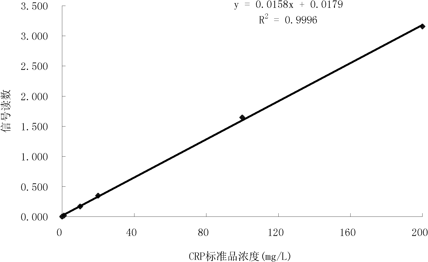
[0056] Example 3 C-reactive protein (CRP) time-resolved fluorescent immunoassay strip

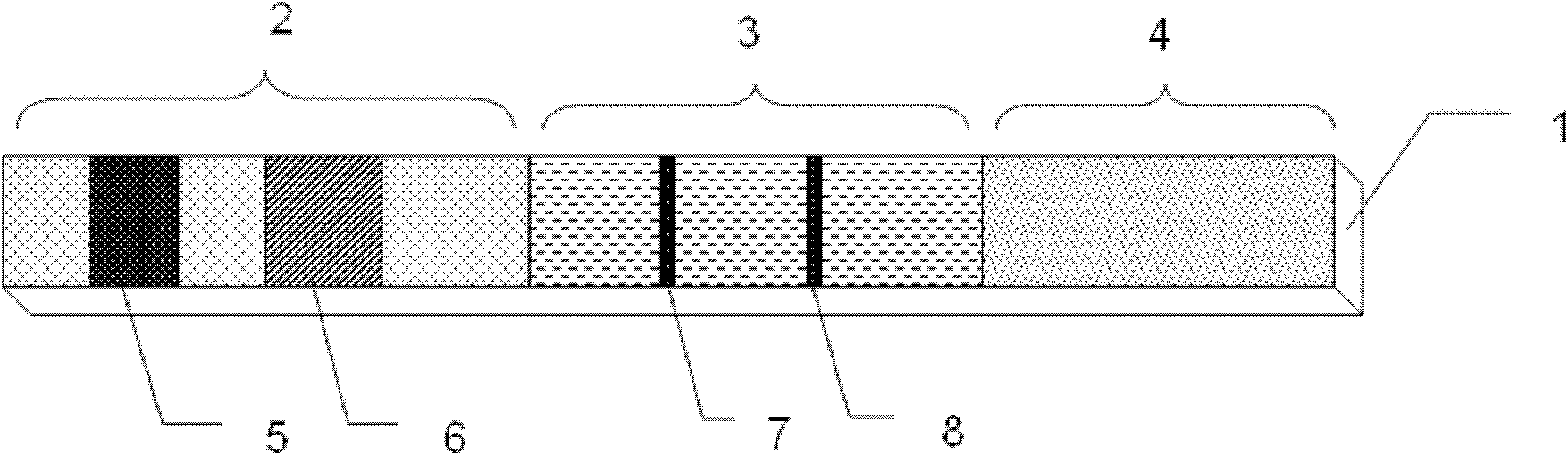
[0057] 1. Preparation of test strip components:

[0058] (1) Preparation of quality control microspheres

[0059] Refer to Example 1 or Example 2 for the preparation method of quality control microspheres.

[0060] (2) Preparation of detection microspheres

[0061] Add 6.4 μl of 20% Tween-20 solution and 2 mg C-reactive protein monoclonal antibody (Boyang Biotechnology (Shanghai) Co., Ltd.) to 10 mg of aldehyde-modified fluorescent microspheres (Boyang Biotechnology (Shanghai) Co., Ltd.) and 16 μl of NaCNBH 3 (25mg / ml, 0.05M pH6.0 MES buffer preparation, ready to use), add 0.1M pH6.0MES to a total volume of 400μl, and incubate at 37° in the dark for 48h. Add 40 μl of Gly solution (75 mg / ml, prepared in 0.05 M MES buffer solution with pH 6.0), and rotate for 2 hours at 37° in the dark. Add 250 μl of N, O-bistrimethylsilylacetamide (200 mg / ml, prepared in 0.05 M MES buffer, pH 6.0) solut...


PUM

PropertyMeasurementUnit
particle diameteraaaaaaaaaa
Sensitivityaaaaaaaaaa

Abstract

The invention discloses a fast time-resolved immunoflourometric chromatographic test strip for quantitative detection as well as a preparation method and application thereof. The test strip comprises three parts including a Fusion 5 film, a cellulose nitrate film and water absorbing paper; by using a double-antibody sandwich method or a competition method principle, a time-resolved fluorescent microsphere is utilized as an immune marker to accurately and fast carry out quantitative detection on an antigen or antibody to be detected.

Description

technical field [0001] The invention relates to the field of biotechnology, in particular to a time-resolved fluorescent immunochromatographic test strip for quantitative detection and a preparation method and application thereof. Background technique [0002] The current rapid detection strips of immunochromatography (lateral flow immunoassay, LFIA) mostly use colloidal gold, colored latex microspheres or fluorescein as labels. The rapid detection products developed based on colloidal gold labeling technology have problems such as low sensitivity, only qualitative or semi-quantitative, and large batch-to-batch differences; although the batch-to-batch difference of colored latex microspheres has improved, the sensitivity is still low. Can be qualitative or semi-quantitative; the sensitivity of immunochromatography based on fluorescein labeling technology has been greatly improved, and quantitative detection can also be performed, but because the sample contains a high fluore...

Claims


Application Information

Patent Timeline
Patent Type & Authority Applications(China)
IPC IPC(8): G01N33/558G01N33/533
Inventor 石晓强马顺华吴茜王海蛟赵卫国
Owner BEYOND DIAGNOSTICS (SHANGHAI) CO LTD
Why Eureka
Try Eureka