Structure and application of Enterovirus 71 3C protease

A protease, enterovirus technology, applied in the field of 3C protease structure, can solve the problem of difficulty in screening specificity

Active Publication Date: 2011-09-28
INST OF MICROBIOLOGY - CHINESE ACAD OF SCI
View PDF1 Cites 8 Cited by
  • Summary
  • Abstract
  • Description
  • Claims
  • Application Information

AI Technical Summary

Problems solved by technology

Therefore, based on the 3C protease structure of HRV or CVB3 virus, there may be a large difference between the modeled EV71-3...

Method used


Image

  • Structure and application of Enterovirus 71 3C protease
  • Structure and application of Enterovirus 71 3C protease
  • Structure and application of Enterovirus 71 3C protease

Examples


Embodiment 1

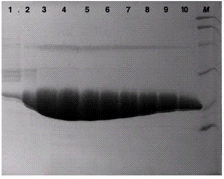
[0032] Example 1 Obtaining of EV71 3C protease

[0033] We successfully expressed the 3C protease of EV71 virus in vitro, and obtained high-purity and active enzyme protein by affinity chromatography and molecular exclusion chromatography.

[0034] The amino acid sequence of EV71 3C protease is shown in SEQ ID NO.1, or the sequence shown in SEQ ID NO.1 is derived from the amino acid sequence of SEQ ID NO.1 through substitution, deletion or addition of one or several amino acids and having 3C protease activity of protein.

[0035] Protein sequence SEQ ID NO.1:

[0036] GPSLDFALSLLRRNIRQVQTDQGHFTMLGVRDRLAVLPRHSQPGKTIWIEHKLVNVLDAVELVDEQGVNLELTLITLDTNEKFRDITKFIPENISTASDA

[0037] 1. Plasmid construction:

[0038] We artificially synthesized the DNA fragment encoding 3C protease, ligated it into the pET-21a vector with Nde1 and Xho1 restriction sites, and inserted a 6-histidine tag (hexa-His-tag) behind the encoding fragment of the enzyme protein. The coding sequence is foll...

Embodiment 2

[0050] Example 2 Structure of EV71 3C protease

[0051] 1. Protein crystallization conditions:

[0052] We used the hang-drop vapor-diffusion method and optimized the conditions to obtain crystals with good diffraction quality. The protein preparation solution used for crystallization was all protein concentration 10mg / ml. The conditions are as follows:

[0053] The crystallization conditions of EV71-3C-C147A: 0.1 M Tris-HCl (pH8.5), 20mM lithium sulfate and 25% (w / v) PEG 5000 MME; the crystal growth of this protein also requires 8mM KPVLRTA polypeptide as an additive.

[0054] The complex crystallization conditions of EV71-3C and AG7088: 0.1 M sodium acetate (pH4.6), 0.2M ammonium sulfate and 25% (w / v) PEG 4000, the molar concentration ratio of protein to compound is 1:3.

[0055] 2. Data collection and structure analysis:

[0056] Data collection uses an indoor X-ray diffractometer (in-house Rigaku MicroMax007 rotating-anode X-ray generator). Data processing uses HKL...

Embodiment 3

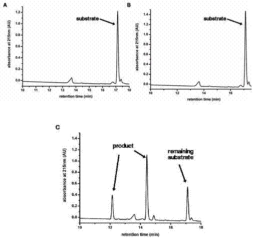
[0060] Example 3 The substrate binding groove of EV71-3C protease

[0061] The nomenclature of each site of the 3C protease substrate binding groove: usually we use the peptide bond cleaved on the substrate polypeptide as the reference point, and the amino acids to the left are named P1, P2, P3, P4,...Pn in sequence, and the amino acids to the right are named in sequence Named P1', P2', P3', P4',...Pn'; and the part of the protease that accommodates these polypeptide amino acids is correspondingly named as the site S1, S2, S3, S4,...Sn, S1 ', S2', S3', S4',...Sn'.

[0062] Through the complex structure of compound AG7088 and EV71-3C protein, we were able to determine the substrate binding groove of EV71-3C protease ( Figure 5 ), Figure 5 The brown stick model in A is AG7088; AG7088 binds in the groove-like structure on the surface of the protease, which is the substrate binding groove of EV71-3C protease (see Figure 5 B). Through structural analysis, we found the amino a...


PUM


Abstract

The invention discloses a structure and application of an Enterovirus 71 (EV71) 3C protease and belongs to the field of RNA virus protein. The invention also discloses an application of the 3C protease and a substrate binding groove thereof in drug design. The protein provided by the invention has a special substrate binding groove and new EV71 virus 3C protease drugs are designed according to the spatial structure of the substrate binding groove, thus the inhibitors aiming at the EV71 virus 3C protease, which are more specific and have better effect, can be obtained, potential drug candidates can be provided for the clinical treatment of hand, foot and mouth disease and the EV71 virus 3C protease has very high application value.

Description

technical field [0001] The invention relates to the structure and application of a 3C protease, in particular to an enterovirus 71 (EV71) 3C protease with a unique substrate binding groove. Background technique [0002] Enterovirus 71 (EV71) is one of the most common pathogens causing HFMD in children. EV71 can also cause infection of the central nervous system, causing aseptic meningitis, acute flaccid paralysis, etc., and even death. In recent years, hand, foot and mouth disease and EV71 infection have exploded worldwide, especially in East Asia (mainland China, Taiwan, Japan, etc.), resulting in huge loss of life, property and economy. At present, there is no effective means of prevention and treatment. [0003] EV71 encodes a virus-specific protease called 3C protease. This protease can process the polyprotein precursor encoded by the virus into functional protein subunits, thus playing an important role in the replication and life cycle of the virus. Humans lack the...

Claims


Application Information

Patent Timeline
IPC IPC(8): C12N9/50G06F19/16C12R1/93
Inventor 高福逯光文
Owner INST OF MICROBIOLOGY - CHINESE ACAD OF SCI
Why Eureka
Try Eureka