Bioelectrochemical sensor for detecting tumor markers and preparation method thereof

A tumor marker, bioelectrochemical technology, applied in the field of new bioelectrochemical sensors and their preparation, to achieve the effect of wide application prospects

Inactive Publication Date: 2011-11-30
SHANGHAI UNIV
View PDF1 Cites 26 Cited by
  • Summary
  • Abstract
  • Description
  • Claims
  • Application Information

AI Technical Summary

Problems solved by technology

However, the detection of a single tumor marker often leads to false positives or false negatives in tumor detection. If multiple tumor markers can be detected at the same time, the sensitivity, specificity and accuracy of tumor detection can be greatly improved.

Method used


Image

  • Bioelectrochemical sensor for detecting tumor markers and preparation method thereof
  • Bioelectrochemical sensor for detecting tumor markers and preparation method thereof
  • Bioelectrochemical sensor for detecting tumor markers and preparation method thereof

Examples


Embodiment 1

[0026] The gold electrode to be treated was sequentially polished on the silk containing alumina (grain size: 1 μm, 0.3 μm, 0.05 μm) mortar, then ultrasonically cleaned with ethanol and ultrapure water for 5 minutes, and then dripped on the surface of the gold electrode 10 μL piranha solution (H 2 o 2 : Concentrated H 2 SO 4 =3:1) for 2 minutes, wash away with ultrapure water, place the gold electrode in 0.5 M sulfuric acid solution, and perform cyclic voltammetry scanning in the voltage range of 0 - 1.6 V, with the scanning speed set at 100 mV / s , about 20 cycles to reach a stable state, and dry it with nitrogen gas to obtain a bare gold electrode with a clean surface, which can be used for the modification of sulfhydryl cDNA. The modification process is: immerse the cleaned gold electrode into the cDNA chain containing 1 μM sulfhydryl group (5'-SH-(CH 2 ) 6 -AGGAT CAACT GCGGC CAGCA CACCC AGATC CT-NH 2 -3') in an Eppendorf tube containing 10 mM Tris-HCl (pH 7.8), 1 mM E...

Embodiment 2

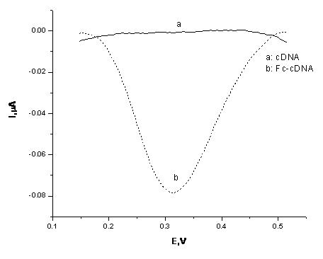
[0028]The gold electrode modified with the cDNA strand was immersed in a mixed solution of 5 mM EDC, 25 mM NHS, and 5 mM ferrocene carboxylic acid, and placed in a water bath at 37°C for 2 hours. During this process, the -NH at the 3' end of the cDNA strand on the gold electrode is modified 2 Condensation reaction occurs with -COOH of ferrocene carboxylic acid, so that Fc is attached to the end of cDNA chain. The result of this modification can be monitored by electrochemical method. like figure 2 In curve a, before Fc modification, because the cDNA chain cannot undergo redox reactions in the voltage range of 0 - 0.6 V, there is no obvious electrical signal; while curve b shows an obvious square wave voltage around 0.31 V An signal, which is generated by the Fc attached to the cDNA, indicates that the Fc molecule was successfully modified to the end of the cDNA chain.

[0029] Example 3: The sample contains only one tumor marker MUC1

Embodiment 3

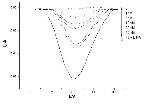
[0030] First, samples containing different concentrations of tumor marker MUC1 (1nM, 5nM, 10nM, 20nM, 40nM) were added to the test solution (10 nM MUC 1 aptamer, 10 nM VEGF 165 aptamer) for 1 hour at room temperature. During this process, part of the MUC1 aptamer in the test solution binds to its target protein, the tumor marker MUC1, and the rest of the MUC1 aptamer and all VEGF 165 The aptamer is freely dissociated in the test solution.

[0031] After the reaction, the gold electrode modified with Fc-cDNA was immersed in 100 μL of the above test solution, and reacted at room temperature for 1.5 hours to measure the electrochemical signal. The result is as image 3 As shown, with the increase of MUC1 concentration, the electrical signal gradually increased, and the reaction was basically saturated when the MUC1 concentration was 20 nM, and the electrical signal increased weakly when the MUC1 concentration was 40 nM. Therefore, we set 20 nM MUC1 as the optimal concentration...


PUM


Abstract

The invention relates to a bioelectrochemical sensor for detecting tumor markers and a preparation method thereof. It is a three-electrode system sensor, which is characterized in that the counter electrode in the three electrodes is a platinum electrode, the reference electrode is a saturated calomel electrode, and the working electrode is a gold electrode, and the gold electrode is decorated with two tumor markers The complementary DNA strand of the DNA aptamer, that is, the cDNA strand, is labeled with an electrochemical probe ferrocene molecule at the end of the cDNA strand. The novel bioelectrochemical sensor for detecting tumor markers of the present invention combines the characteristics of highly specific binding of DNA aptamers to target proteins and high sensitivity of electrochemical detection methods, making early diagnosis of cancer possible.

Description

technical field [0001] The invention relates to a novel bioelectrochemical sensor and a preparation method thereof, in particular to a bioelectrochemical sensor for detecting tumor markers and a preparation method thereof. Background of the invention [0002] Cancer is one of the diseases with high morbidity and mortality that seriously threatens human health. However, if cancer can be diagnosed at an early stage of development, the survival rate of patients will be greatly improved. It can be said that early diagnosis and early treatment are one of the most effective ways to prevent cancer development and reduce mortality. Tumor markers are a class of substances that are produced, secreted, and released into blood, cells, and body fluids by tumor cells during tumorigenesis and proliferation, reflecting the existence and growth of tumors. It is a specific representation of the state of tumor cells in the human body, and can directly reflect the development of tumor cells ...

Claims


Application Information

Patent Timeline
Patent Type & Authority Applications(China)
IPC IPC(8): G01N27/406G01N27/327C12N15/11
Inventor 李根喜赵婧郭超赵洪喜吴瑶陈阳阳
Owner SHANGHAI UNIV
Why Eureka
Try Eureka