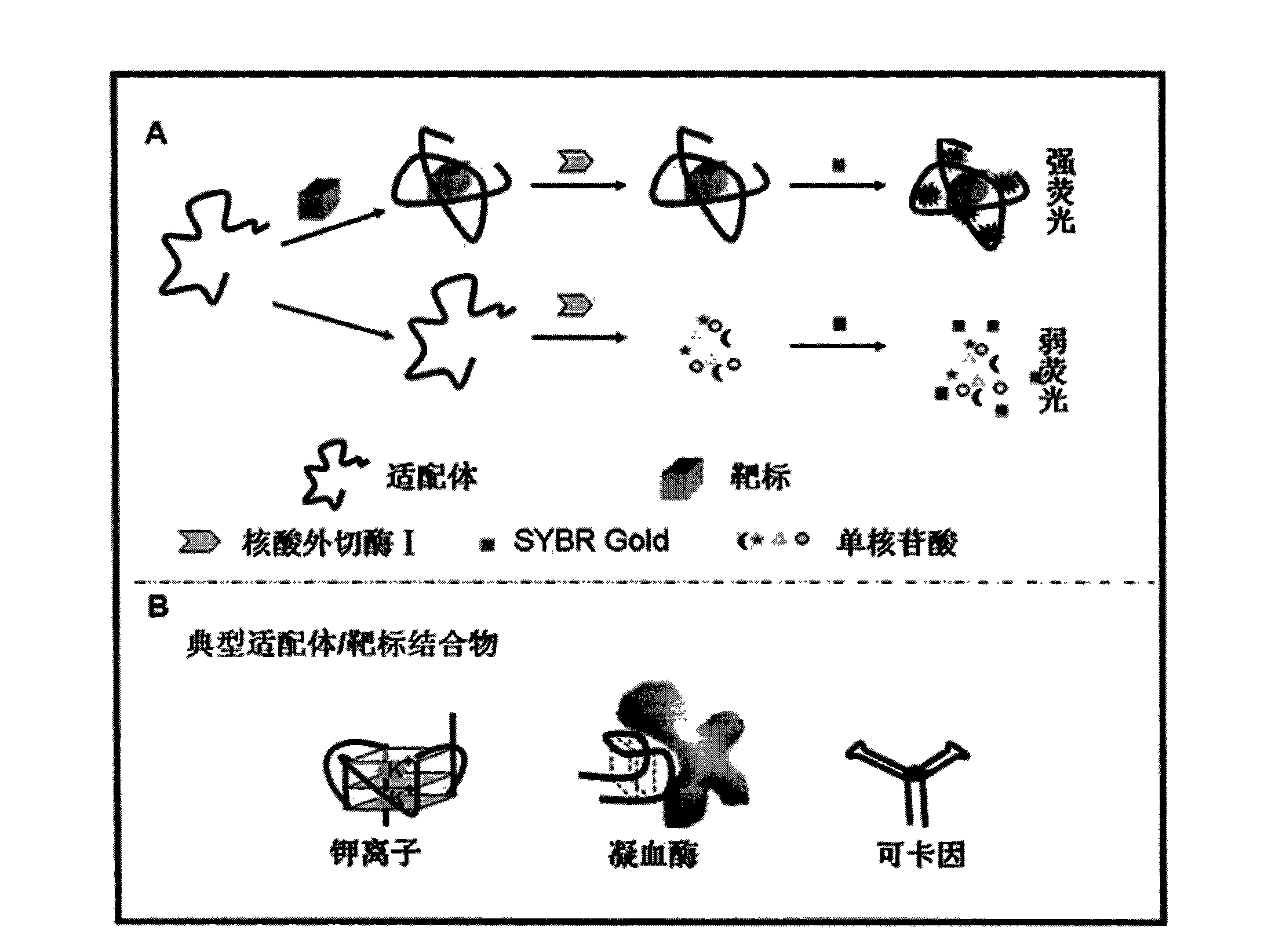
Structure-switching aptamer based highly-sensitive unmarked fluorescent detection method
A fluorescence detection and aptamer technology, which is applied in the determination/inspection of microorganisms, biochemical equipment and methods, etc., to achieve the effects of high detection specificity, resistance to interference from analogues and interferents, and low cost
- Summary
- Abstract
- Description
- Claims
- Application Information
AI Technical Summary
Problems solved by technology
Method used
Image
Examples
Embodiment 1
[0033] Example 1 uses probe 1 to detect different concentrations of K +
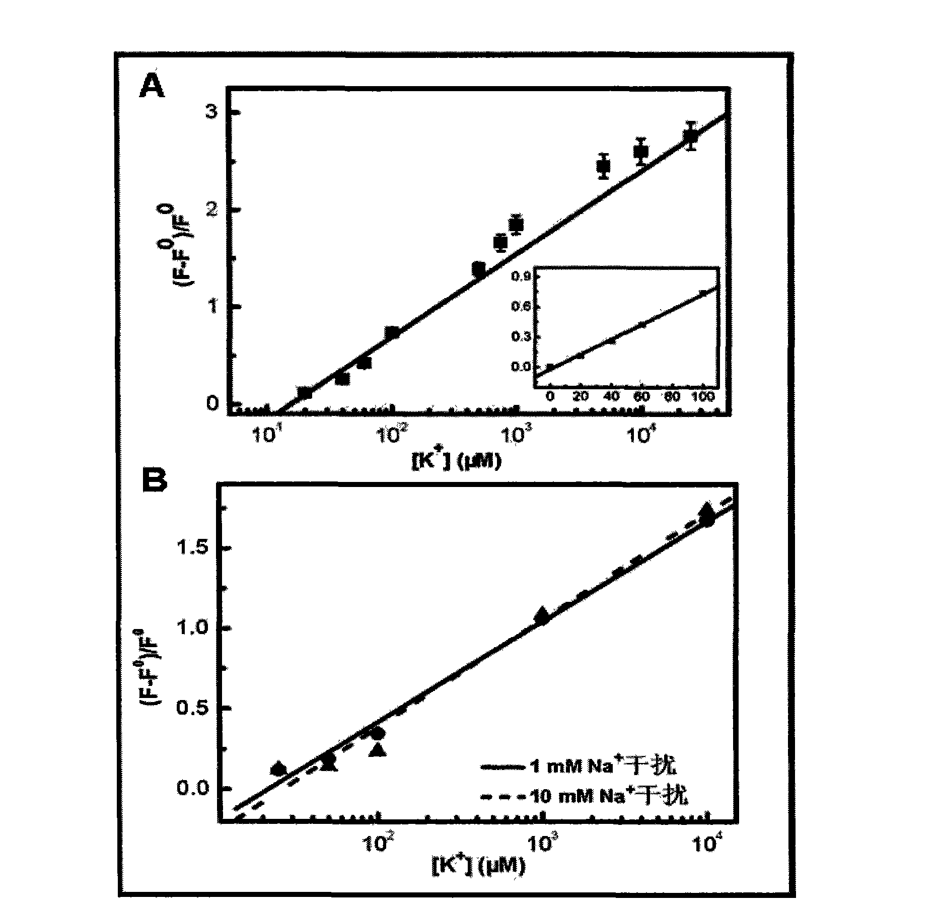
[0034] 50mM Tris-HCl, 2mM MgCl 2 , pH 8.3 Probe 1 was diluted to a concentration of 10 μM, heated at 90 °C for 5 min, and then slowly cooled to room temperature. Simultaneously with 50mM Tris-HCl, 2mM MgCl 2 , pH 8.3 diluted to prepare different concentrations of K + . Take 10 μL of probe 1 solution and 969 μL of different concentrations of K + The solution was mixed evenly in a centrifuge tube, and reacted for 12 hours at room temperature. Add 5U Exo I respectively, react at 37°C for 40min, and then heat at 80°C for 15min to stop the enzyme cleavage reaction. Then add 20μL of 50mM Tris-HCl, 2mM MgCl 2 , pH 8.3 diluted 10X SYBR Gold solution was mixed evenly, and stained for 20min. Scan and analyze the results with an atomic fluorescence spectrometer F-4500 from Shanghai Hitachi figure 2 (A).
[0035] The results show that with K + As the concentration increases, the fluorescence intensity in...
Embodiment 2
[0036] Example 2. Containing Na + Detection of K in the buffer + , examine Na + to K + detection interference.
[0037] 50mM Tris-HCl, 2mM MgCl 2 , 1 mM NaCl, pH 8.3 Probe 1 was diluted to a concentration of 10 μM, heated at 90 °C for 5 min, and then slowly cooled to room temperature. Simultaneously with 50 mM Tris-HCl, 2mM MgCl 2 , 1mM NaCl, pH 8.3 by diluting to prepare different concentrations of K + . Take 10 μL of probe 1 solution and 969 μL of different concentrations of K + The solution was mixed evenly in a centrifuge tube, and reacted for 12 hours at room temperature. Add 5U Exo I respectively, react at 37°C for 40min, and then heat at 80°C for 15min to stop the enzyme cleavage reaction. Then add 20μL of 50mM Tris-HCl, 2mM MgCl 2 , 1mM NaCl, pH 8.3 diluted 10X SYBR Gold solution was mixed evenly, and stained for 20min. Scan and analyze the results with an atomic fluorescence spectrometer F-4500 from Shanghai Hitachi figure 2 Solid line in (B).
[0038] 5...
Embodiment 3
[0040] Embodiment 3: investigate this fluorescence method to K + Specificity of detection.
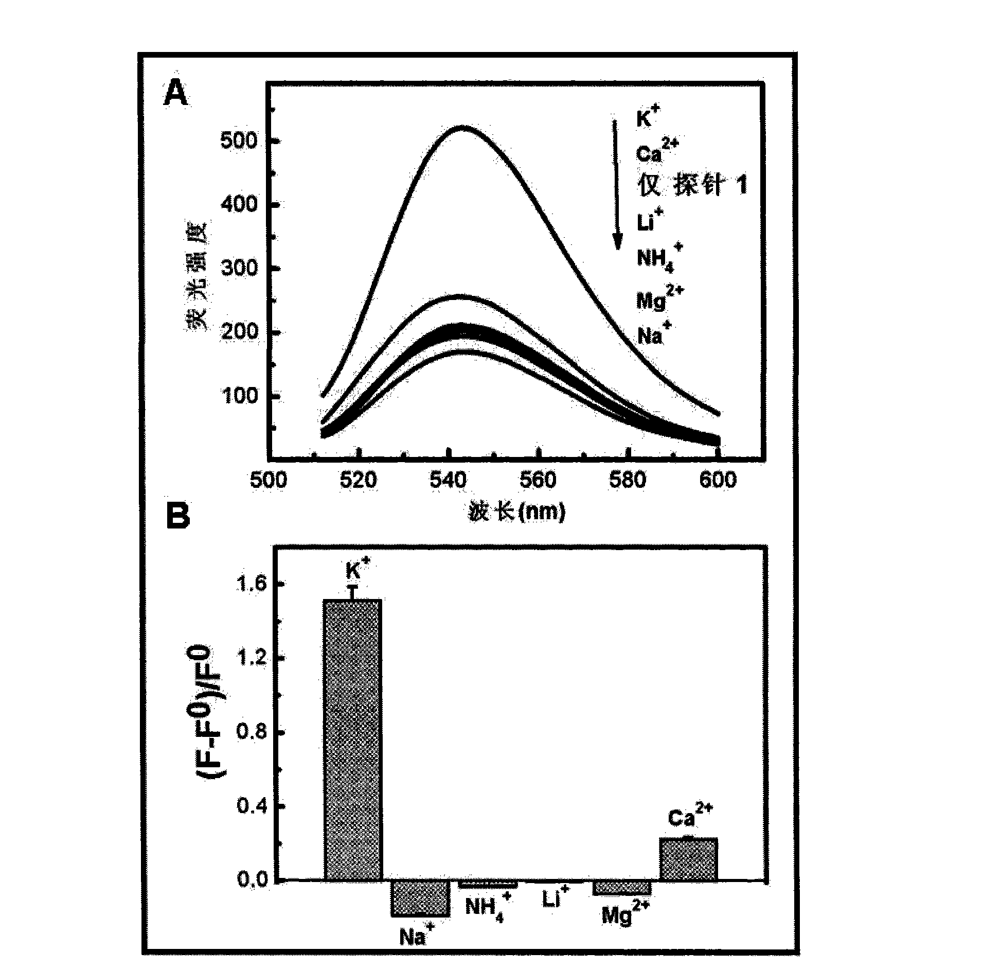
[0041] 50mM Tris-HCl, 2mM MgCl 2 , pH 8.3 Probe 1 was diluted to a concentration of 10 μM, heated at 90 °C for 5 min, and then slowly cooled to room temperature. Simultaneously with 50mM Tris-HCl, 2mM MgCl 2 , pH 8.3 by diluting to prepare 1 mM of the different ions. Take 10 μL of probe 1 solution and 969 μL of solutions of different ions and mix them evenly in a centrifuge tube, and react at room temperature for 12 hours. Add 5U Exo I respectively, react at 37°C for 40min, and then heat at 80°C for 15min to stop the enzyme cleavage reaction. Then add 20μL of 50mM Tris-HCl, 2mM MgCl 2 , pH 8.3 diluted 10X SYBR Gold solution was mixed evenly, and stained for 20min. The atomic fluorescence photometer F-4500 of Shanghai Hitachi company was used to scan and analyze the results as follows: image 3 (A)-(B) shown.
[0042] The results show that the common cations on K + Detection in...
PUM
 Abstract
Description
Claims
Application Information
 - R&D
- Intellectual Property
- Life Sciences
- Materials
- Tech Scout
- Unparalleled Data Quality
- Higher Quality Content
- 60% Fewer Hallucinations
Browse by: Latest US Patents, China's latest patents, Technical Efficacy Thesaurus, Application Domain, Technology Topic, Popular Technical Reports.
© 2025 PatSnap. All rights reserved.Legal|Privacy policy|Modern Slavery Act Transparency Statement|Sitemap|About US| Contact US: help@patsnap.com


