
Use of CD133 in preparation of tumor marker and kit of CD133

A tumor marker and kit technology, applied in the field of molecular biology and tumor drugs, can solve problems such as no discovery effect

Inactive Publication Date: 2012-09-26
FUDAN UNIV
View PDF4 Cites 2 Cited by
  • Summary
  • Abstract
  • Description
  • Claims
  • Application Information

AI Technical Summary

Problems solved by technology

In tumor studies, it has been found that CD133-positive tumor cells have stronger self-renewal and proliferation capabilities than CD133-negative cells, but their role in glioma invasion has not been found so far

Method used


Image

  • Use of CD133 in preparation of tumor marker and kit of CD133
  • Use of CD133 in preparation of tumor marker and kit of CD133
  • Use of CD133 in preparation of tumor marker and kit of CD133

Examples


Embodiment 1

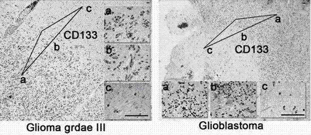
[0057] The C57 mouse orthotopic model of GL261 was constructed with luciferase-labeled GL261 mouse glioma cells, and the tumor growth was dynamically detected by live imaging of small animals. Using mouse glioma model and human malignant glioma specimens, CD133 (CD133-Flag sequence is shown in SEQ ID NO 1, and its luciferase sequence is shown in SEQ ID NO 2) was detected by immunohistochemistry in glioma, glial The junction of glioma and normal tissue and the expression characteristics and differences in normal tissue. The results showed that the glioma invaded the normal brain tissue area obviously. CD133 immunohistochemical staining of brain slices showed that CD133 was highly expressed in the tumor invasion area, while CD133 was less expressed in the tumor and normal brain tissue. The same results were also verified in human glioma specimens. CD133 immunohistochemical staining of 18 glioma specimens with junctional tumor and normal tissue showed that the expression of CD1...

Embodiment 2

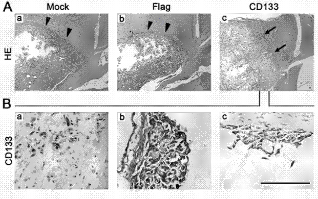
[0061] A plasmid expressing CD133 was constructed, and CD133 was overexpressed in the above two glioma cells. Using the C57 mouse model of GL261 and the U87 nude mouse model, the border of tumor formation, the number of daughter tumors, the invasion of the corpus callosum and the spread of the ventricles were observed after CD133 overexpression. The results showed that the tumors formed in the Mock and Flag groups had smooth edges, while the tumors in the CD133 group showed high invasiveness, showing obvious invasion to normal brain tissue, and a large amount of cerebrospinal fluid dissemination and planting. These disseminated foci were all CD133+, suggesting that they were all derived from exogenous CD133 cells. The same phenomenon as above was observed in the U87-GFP and U87-CD133-GFP nude mouse models, that is, the tumor invasiveness of the CD133 group was significantly stronger than that of the Flag group, and a large number of daughter tumors were formed in the CD133 gro...

Embodiment 3

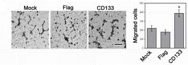
[0065] Using GL261 and U87 glioma cells overexpressing CD133, the expression of CD133 was further analyzed by Transwell model, flow cytometry detection of cell cycle, gelatin zymography test, cell adhesion test, in vitro scratch test and cytoskeleton protein F-actin staining. Role in glioma invasion.

[0066] The results of Transwell invasion assay showed that the number of invasive cells in the CD133 group was significantly more than that in the Mock and Flag groups, suggesting that CD133 can significantly promote the invasion of GL261 glioma cells in vitro. There was no statistical difference in the cell cycle distribution of the three groups of cells, which ruled out the influence of the cell cycle on the results of the Transwell invasion assay. The results of MMP zymography experiments indicated that there was no difference in the activities of MMP-2 and MMP-9 among the three groups of cells, indicating that CD133 had no significant effect on the ability of glioma cells to d...


PUM


Abstract

The invention relates to the fields of tumor molecular biology and tumor drug treatment. The invention provides a use of CD133 as a tumor cell invasion marker and an application with CD133 as a target for screening drugs that inhibit tumor cell invasion. According to the invention, technologies of plasmid transfection, rat glioma model establishment, vivo imaging, immunohistochemistry and Transwellare employed to verify the high expression of the CD133 in invasion foci around glioma, and it is verified that highly-expressed glioma cells are more susceptible to invasion in vivo and in vitro. Further research indicates thatthe CD133 makes glioma more susceptible to invasion by improving tumor cell migration capability and altering cytoskeletal. The CD133 can provide tumor treatment programme selection and antitumor drug screening with new ideas.

Description

technical field [0001] The invention relates to the fields of molecular biology and tumor medicine. Specifically, the present invention relates to the role of a tumor stem cell surface marker CD133 in glioma invasion, and the potential application of CD133 and downstream pathways in drug screening and treatment of tumors. Background technique [0002] Brain astroglioma is a fatal and common intracranial tumor. Under the premise of conventional surgical treatment, radiotherapy and chemotherapy, the average survival time of patients is about 15 months. The reason why glioma is difficult to cure is that glioma cells are easy to invade and spread to the surrounding normal brain tissue, resulting in incomplete surgical resection and tumor recurrence. However, the current understanding of the mechanism and characteristics of glioma invasion is still insufficient. Therefore, exploring the molecular mechanism of glioma invasion is the key to the treatment of glioma. [0003] Exis...

Claims


Application Information

Patent Timeline
IPC IPC(8): G01N33/68
Inventor 江建海魏湲颜邹飞江一舟许诺王杉杉
Owner FUDAN UNIV
Learn More